1IIB image
Deposition Date 1996-12-23
Release Date 1997-12-24
Last Version Date 2024-02-07
Entry Detail
PDB ID:
1IIB
Title:
CRYSTAL STRUCTURE OF IIBCELLOBIOSE FROM ESCHERICHIA COLI
Biological Source:
Source Organism:
Method Details:
Experimental Method:
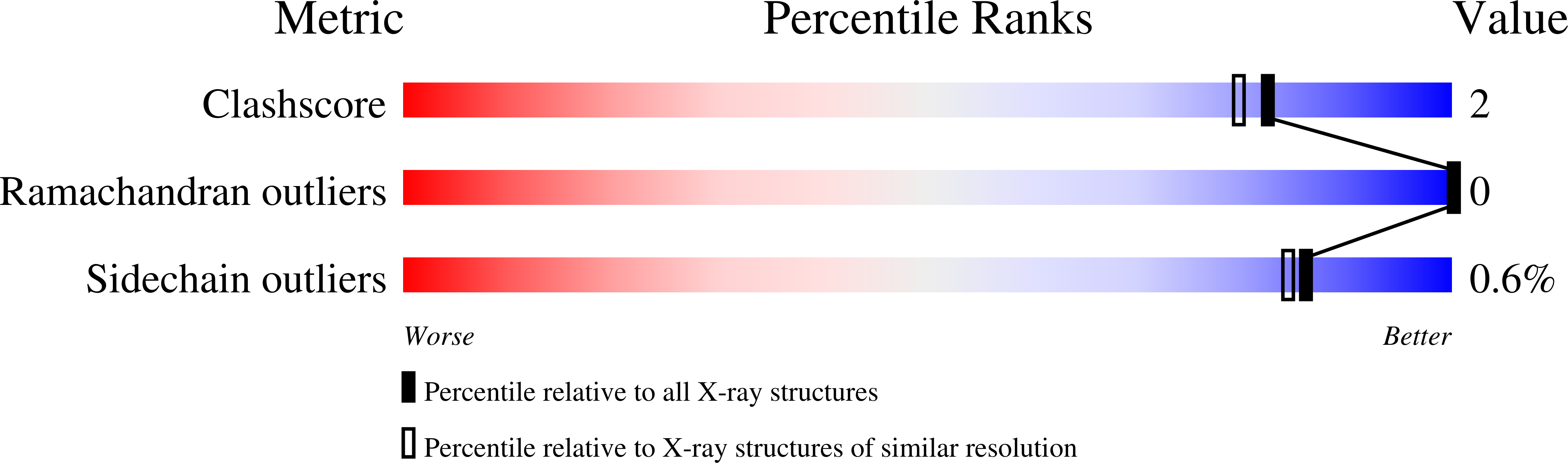
Resolution:
1.80 Å
R-Value Free:
0.24
R-Value Work:
0.18
R-Value Observed:
0.18
Space Group:
P 1 21 1
Macromolecular Entities
Structures with similar UniProt ID
Protein Blast
Polymer Type:polypeptide(L)
Molecule:ENZYME IIB OF THE CELLOBIOSE-SPECIFIC PHOSPHOTRANSFERASE SYSTEM
Gene (Uniprot):chbB
Mutagens:C10S
Chain IDs:A, B
Chain Length:106
Number of Molecules:2
Biological Source:Escherichia coli
Primary Citation
The structure of an energy-coupling protein from bacteria, IIBcellobiose, reveals similarity to eukaryotic protein tyrosine phosphatases.
Structure 5 217 225 (1997)
PMID: 9032081 DOI: 10.1016/S0969-2126(97)00180-9

Abstact

BACKGROUND . The bacterial phosphoenolpyruvate-dependent phosphotransferase system (PTS) mediates the energy-driven uptake of carbohydrates and their concomitant phosphorylation. In addition, the PTS is intimately involved in the regulation of a variety of metabolic and transcriptional processes in the bacterium. The multiprotein PTS consists of a membrane channel and at least four cytoplasmic proteins or protein domains that sequentially transfer a phosphoryl group from phosphoenolpyruvate to the transported carbohydrate. Determination of the three-dimensional structure of the IIB enzymes within the multiprotein complex would provide insights into the mechanisms by which they promote efficient transport by the membrane channel IIC protein and phosphorylate the transported carbohydrate on the inside of the cell. RESULTS . The crystal structure of the IIB enzyme specific for cellobiose, IIBcellobiose (molecular weight 11.4 kDa), has been determined to a resolution of 1.8 and refined to an R factor of 18.7% (Rfree of 24. 1%). The enzyme consists of a single four-stranded parallel beta sheet flanked by helices on both sides. The phosphorylation site (Cys 10) is located at the C-terminal end of the first beta strand. No positively charged residues, which could assist in phosphoryl-transfer, can be found in or near the active site. The fold of IIBcellobiose is remarkably similar to that of the mammalian low molecular weight protein tyrosine phosphatases. CONCLUSIONS . A comparison between IIBcellobiose and the structurally similar low molecular weight protein tyrosine phosphatases provides insight into the mechanism of the phosphoryltransfer reactions in which IIBcellobiose is involved. The differences in tertiary structure and active-site composition between IIBcellobiose and the glucose-specific IIBglucose give a structural explanation why the carbo-hydrate-specific components of different families cannot complement each other.

Legend

Protein

Chemical

Disease

Primary Citation of related structures