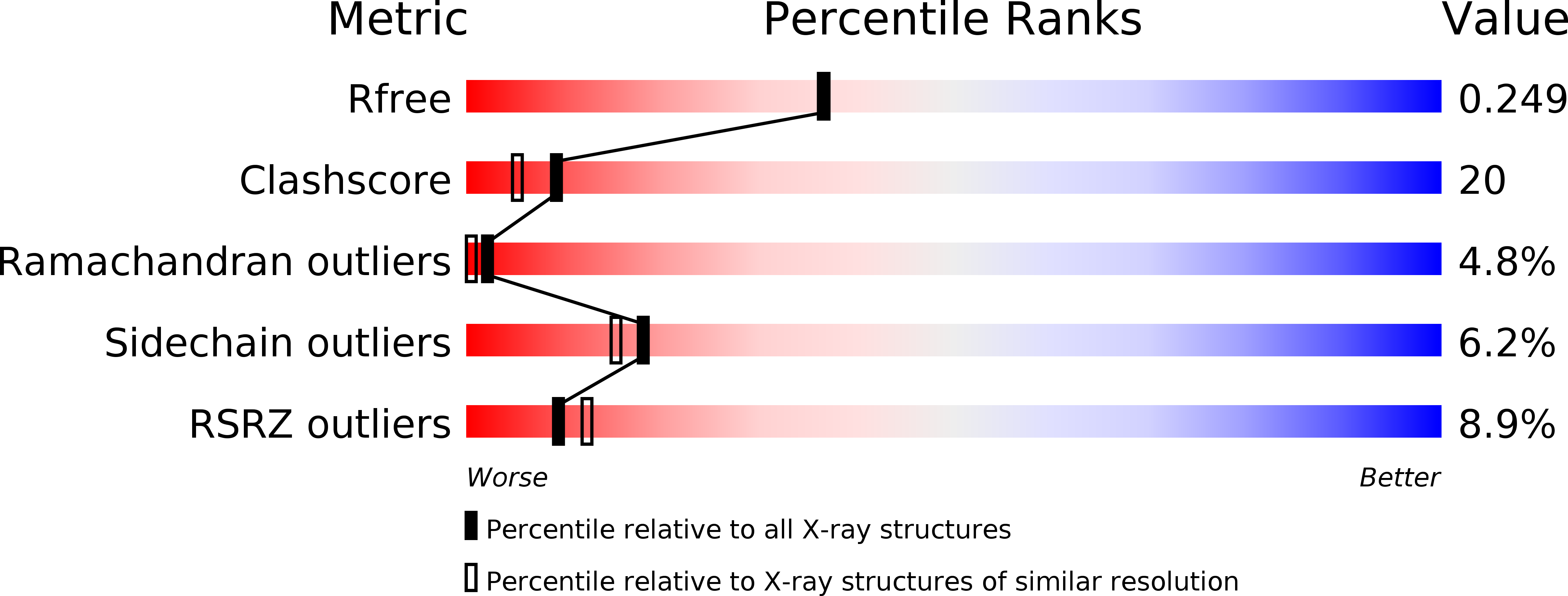
1II6 image
Deposition Date 2001-04-20
Release Date 2001-07-18
Last Version Date 2024-04-03
Entry Detail
PDB ID:
1II6
Keywords:
Title:
Crystal Structure of the Mitotic Kinesin Eg5 in Complex with Mg-ADP.
Biological Source:
Source Organism:
Homo sapiens (Taxon ID: 9606)
Method Details:
Experimental Method:
Resolution:
2.10 Å
R-Value Free:
0.25
R-Value Work:
0.21
Space Group:
P 1 21 1
Macromolecular Entities
Polymer Type:polypeptide(L)
Molecule:KINESIN-RELATED MOTOR PROTEIN Eg5
Gene (Uniprot):KIF11
Chain IDs:A, B
Chain Length:368
Number of Molecules:2
Biological Source:Homo sapiens
Primary Citation
Crystal structure of the mitotic spindle kinesin Eg5 reveals a novel conformation of the neck-linker.
J.Biol.Chem. 276 25496 25502 (2001)
PMID: 11328809 DOI: 10.1074/jbc.M100395200

Abstact

Success of mitosis depends upon the coordinated and regulated activity of many cellular factors, including kinesin motor proteins, which are required for the assembly and function of the mitotic spindle. Eg5 is a kinesin implicated in the formation of the bipolar spindle and its movement prior to and during anaphase. We have determined the crystal structure of the Eg5 motor domain with ADP-Mg bound. This structure revealed a new intramolecular binding site of the neck-linker. In other kinesins, the neck-linker has been shown to be a critical mechanical element for force generation. The neck-linker of conventional kinesin is believed to undergo an ordered-to-disordered transition as it translocates along a microtubule. The structure of Eg5 showed an ordered neck-linker conformation in a position never observed previously. The docking of the neck-linker relies upon residues conserved only in the Eg5 subfamily of kinesin motors. Based on this new information, we suggest that the neck-linker of Eg5 may undergo an ordered-to-ordered transition during force production. This ratchet-like mechanism is consistent with the biological activity of Eg5.

Legend

Protein

Chemical

Disease

Primary Citation of related structures