Deposition Date
2001-04-20
Release Date
2001-11-21
Last Version Date
2023-08-16
Entry Detail
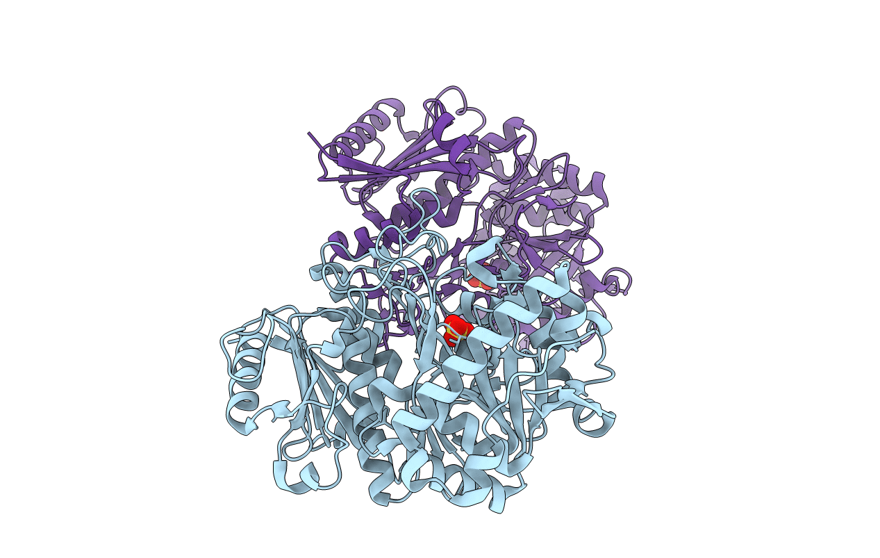
PDB ID:
1II2
Keywords:
Title:
Crystal Structure of Phosphoenolpyruvate Carboxykinase (PEPCK) from Trypanosoma cruzi
Biological Source:
Source Organism(s):
Trypanosoma cruzi (Taxon ID: 5693)
Expression System(s):
Method Details:
Experimental Method:
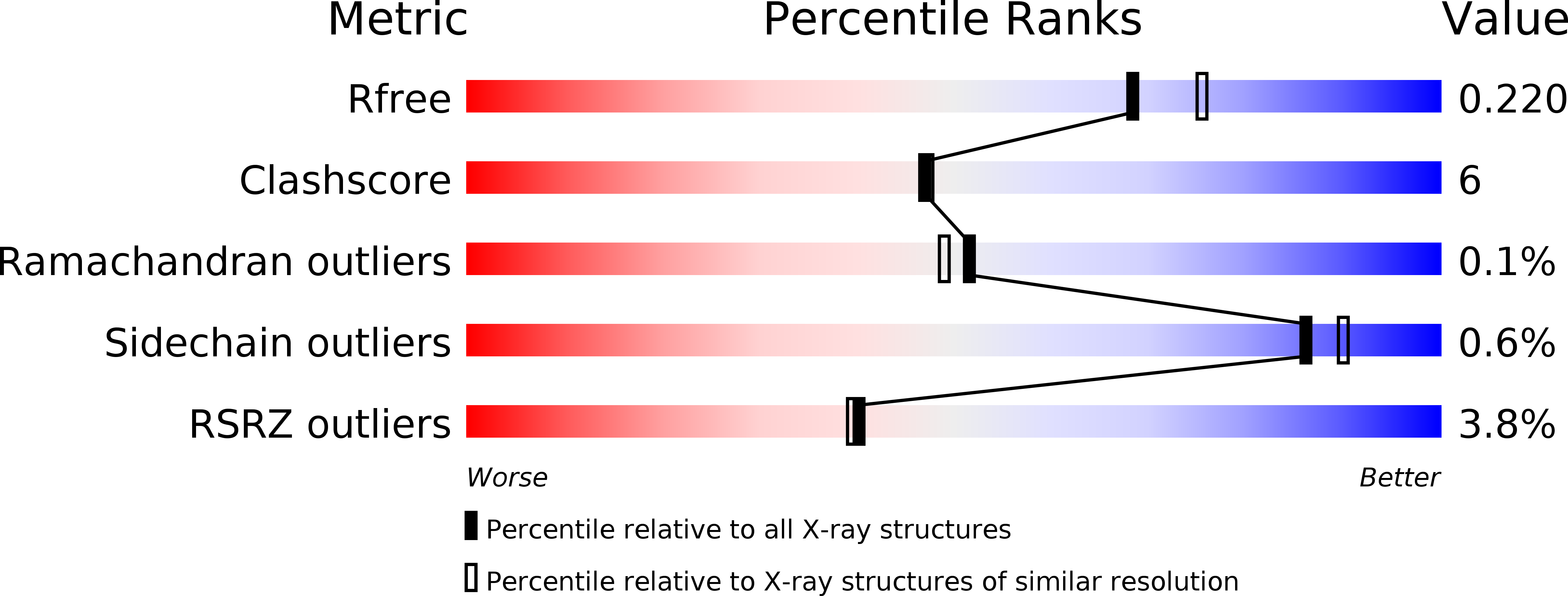
Resolution:
2.00 Å
R-Value Free:
0.22
R-Value Work:
0.19
R-Value Observed:
0.19
Space Group:
P 21 21 21