1IHR image
Deposition Date 2001-04-20
Release Date 2001-08-01
Last Version Date 2024-02-07
Entry Detail
PDB ID:
1IHR
Title:
Crystal structure of the dimeric C-terminal domain of TonB
Biological Source:
Source Organism:
Escherichia coli (Taxon ID: 562)
Host Organism:
Method Details:
Experimental Method:
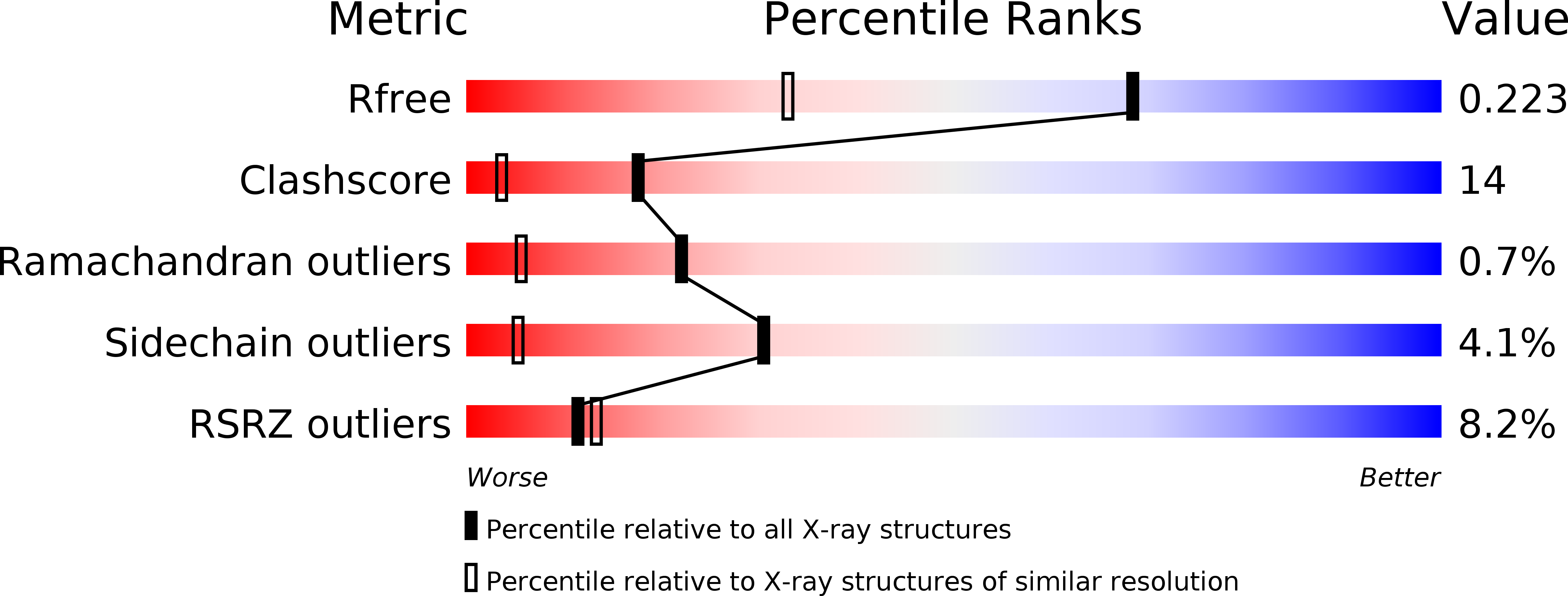
Resolution:
1.55 Å
R-Value Free:
0.23
R-Value Work:
0.15
R-Value Observed:
0.16
Space Group:
P 21 21 2
Macromolecular Entities
Structures with similar UniProt ID
Protein Blast
Polymer Type:polypeptide(L)
Molecule:TonB protein
Gene (Uniprot):tonB
Chain IDs:A, B
Chain Length:76
Number of Molecules:2
Biological Source:Escherichia coli
Ligand Molecules
Primary Citation
Crystal structure of the dimeric C-terminal domain of TonB reveals a novel fold.
J.Biol.Chem. 276 27535 27540 (2001)
PMID: 11328822 DOI: 10.1074/jbc.M102778200

Abstact

The TonB-dependent complex of Gram-negative bacteria couples the inner membrane proton motive force to the active transport of iron.siderophore and vitamin B(12) across the outer membrane. The structural basis of that process has not been described so far in full detail. The crystal structure of the C-terminal domain of TonB from Escherichia coli has now been solved by multiwavelength anomalous diffraction and refined at 1.55-A resolution, providing the first evidence that this region of TonB (residues 164-239) dimerizes. Moreover, the structure shows a novel architecture that has no structural homologs among any known proteins. The dimer of the C-terminal domain of TonB is cylinder-shaped with a length of 65 A and a diameter of 25 A. Each monomer contains three beta strands and a single alpha helix. The two monomers are intertwined with each other, and all six beta-strands of the dimer make a large antiparallel beta-sheet. We propose a plausible model of binding of TonB to FhuA and FepA, two TonB-dependent outer-membrane receptors.

Legend

Protein

Chemical

Disease

Primary Citation of related structures