
Deposition Date
2001-04-19
Release Date
2001-05-02
Last Version Date
2024-02-07
Entry Detail
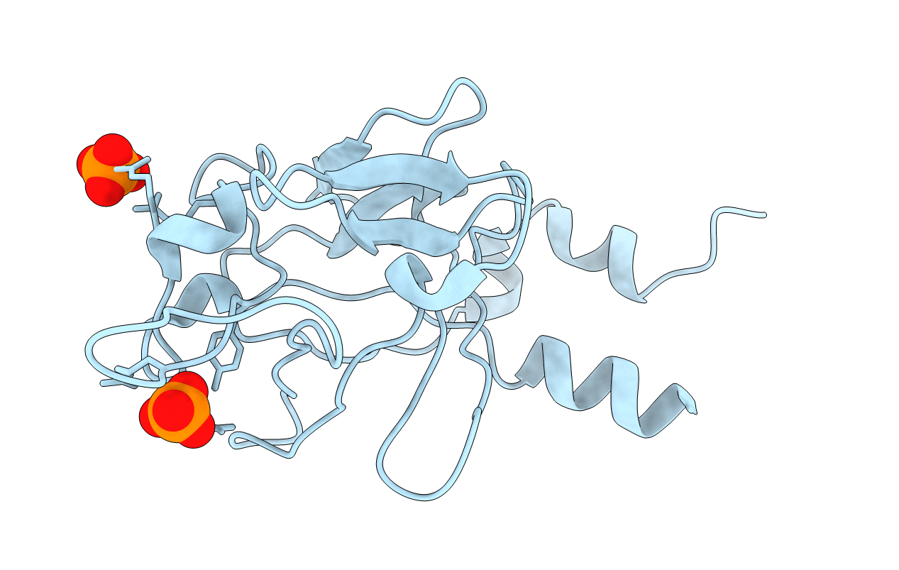
PDB ID:
1IHK
Keywords:
Title:
CRYSTAL STRUCTURE OF FIBROBLAST GROWTH FACTOR 9 (FGF9)
Biological Source:
Source Organism(s):
Homo sapiens (Taxon ID: 9606)
Expression System(s):
Method Details:
Experimental Method:
Resolution:
2.20 Å
R-Value Free:
0.22
R-Value Work:
0.20
R-Value Observed:
0.20
Space Group:
I 41 2 2
