1IHB image
Deposition Date 1997-10-25
Release Date 1998-12-02
Last Version Date 2024-02-07
Entry Detail
PDB ID:
1IHB
Title:
CRYSTAL STRUCTURE OF P18-INK4C(INK6)
Biological Source:
Source Organism:
Homo sapiens (Taxon ID: 9606)
Method Details:
Experimental Method:
Resolution:
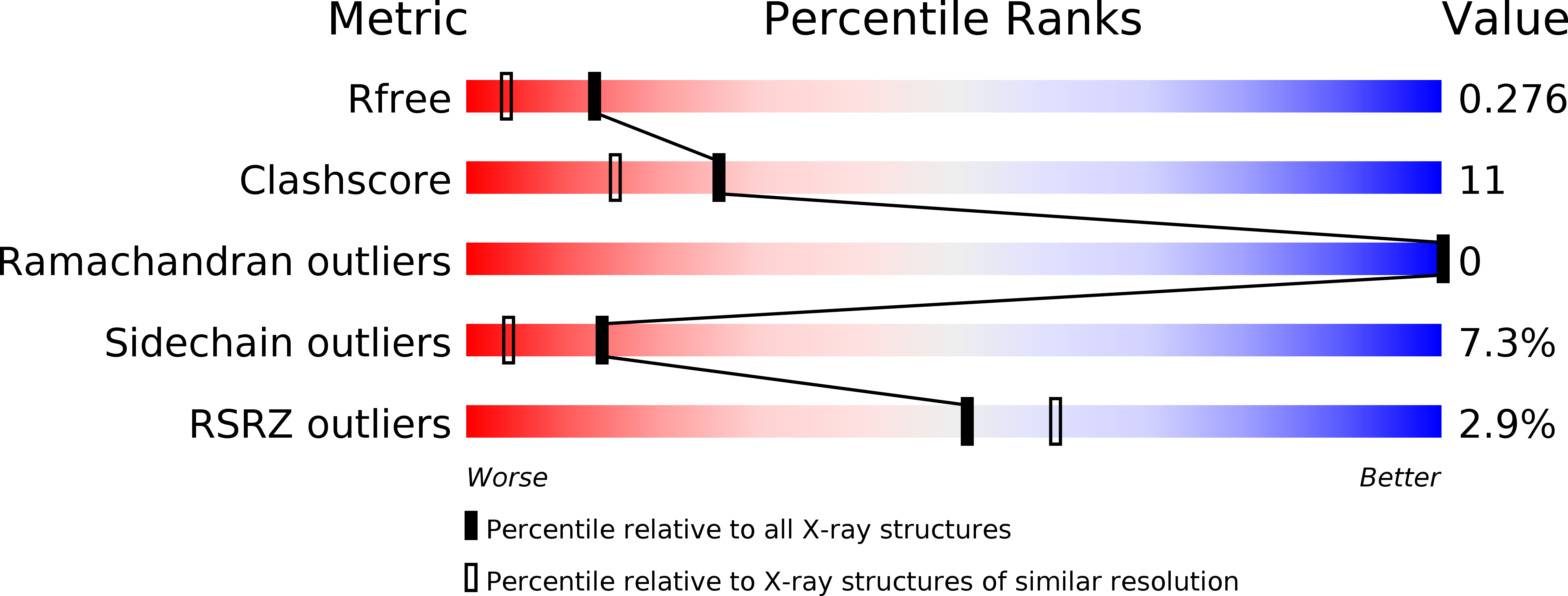
1.95 Å
R-Value Free:
0.28
R-Value Work:
0.20
R-Value Observed:
0.20
Space Group:
P 21 21 2
Macromolecular Entities
Polymer Type:polypeptide(L)
Molecule:CYCLIN-DEPENDENT KINASE 6 INHIBITOR
Gene (Uniprot):CDKN2C
Chain IDs:A, B
Chain Length:162
Number of Molecules:2
Biological Source:Homo sapiens
Primary Citation
Crystal structure of the CDK4/6 inhibitory protein p18INK4c provides insights into ankyrin-like repeat structure/function and tumor-derived p16INK4 mutations.
Nat.Struct.Biol. 5 74 81 (1998)
PMID: 9437433 DOI: 10.1038/nsb0198-74

Abstact

p18INK4c is a member of a family of INK4 proteins that function to arrest the G1 to S cell cycle transition by inhibiting the activity of the cyclin-dependent kinases 4 and 6. The X-ray crystal structure of the human p18INK4c protein to a resolution of 1.95 A reveals an elongated molecule comprised of five contiguous 32- or 33-residue ankyrin-like repeat units. Each ankyrin-like repeat contains a beta-strand helix-turn-helix extended strand beta-strand motif that associates with neighboring motifs through beta-sheet, and helical bundle interactions. Conserved ankyrin-like repeat residues function to facilitate the ankyrin repeat fold and the tertiary interactions between neighboring repeat units. A large percentage of residues that are conserved among INK4 proteins and that map to positions of tumor-derived p16INK4 mutations play important roles in protein stability. A subset of these residues suggest an INK4 binding surface for the cyclin-dependent kinases 4 and 6. This surface is centered around a region that shows structural features uncharacteristic of ankyrin-like repeat units.

Legend

Protein

Chemical

Disease

Primary Citation of related structures