1IGM image
Deposition Date 1992-07-10
Release Date 1993-10-31
Last Version Date 2024-10-23
Entry Detail
PDB ID:
1IGM
Keywords:
Title:
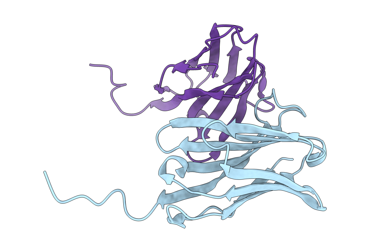
THREE DIMENSIONAL STRUCTURE OF AN FV FROM A HUMAN IGM IMMUNOGLOBULIN
Biological Source:
Source Organism:
Homo sapiens (Taxon ID: 9606)
Method Details:
Experimental Method:
Resolution:
2.30 Å
R-Value Observed:
0.20
Space Group:
P 43 21 2
Macromolecular Entities
Polymer Type:polypeptide(L)
Molecule:IGM-KAPPA POT FV (HEAVY CHAIN)
Chain IDs:B (auth: H)
Chain Length:129
Number of Molecules:1
Biological Source:Homo sapiens
Polymer Type:polypeptide(L)
Molecule:IGM-KAPPA POT FV (LIGHT CHAIN)
Chain IDs:A (auth: L)
Chain Length:115
Number of Molecules:1
Biological Source:Homo sapiens
Primary Citation
Three-dimensional structure of an Fv from a human IgM immunoglobulin.
J.Mol.Biol. 228 188 207 (1992)
PMID: 1447781 DOI: 10.1016/0022-2836(92)90500-J

Abstact

An IgM(kappa) immunoglobulin from a patient (Pot) with Waldenstrom's macroglobulinemia was hydrolyzed with pepsin to release a fragment consisting of the 'variable' (V) domains of the light and heavy chains plus eight residue 'tails' from the 'constant' (C) domains. The crystal structure of this fragment was determined at 2.3 A resolution by molecular replacement and crystallographic refinement methods. When examined separately, the light chain component closely resembles another human kappa chain (Rei) in both the beta-pleated sheet regions and the 'hypervariable' loops. The conserved pleated sheets in the heavy chain are similar to those in the human Kol IgG1 protein, but the third hypervariable loop in particular is different from that in any immunoglobulin structure described to date. As in the Kol protein, this loop blocks the access to any internal active site along the light-heavy chain interface. Unlike the Kol protein, however, the loop does not protrude beyond the boundaries of a conventional antigen combining site. Instead, it forms a very compact structure, which fills almost all residual space between the domains. This is an example of one dominant complementarity-determining region (CDR) essentially negating the diversity possible with five other CDRs in the two chains. Ordered water molecules are associated with light chain constituents along the interface, but not with CDR3 of the heavy chain. In screening exercises the Pot IgM failed to bind a wide variety of peptides. Together, the results suggest that ligand binding can only occur on external surfaces of the protein. These surfaces carry a limited number of side chains usually assigned to CDRs in more typical antibodies.

Legend

Protein

Chemical

Disease

Primary Citation of related structures
Feedback Form
Name
Email
Institute
Feedback