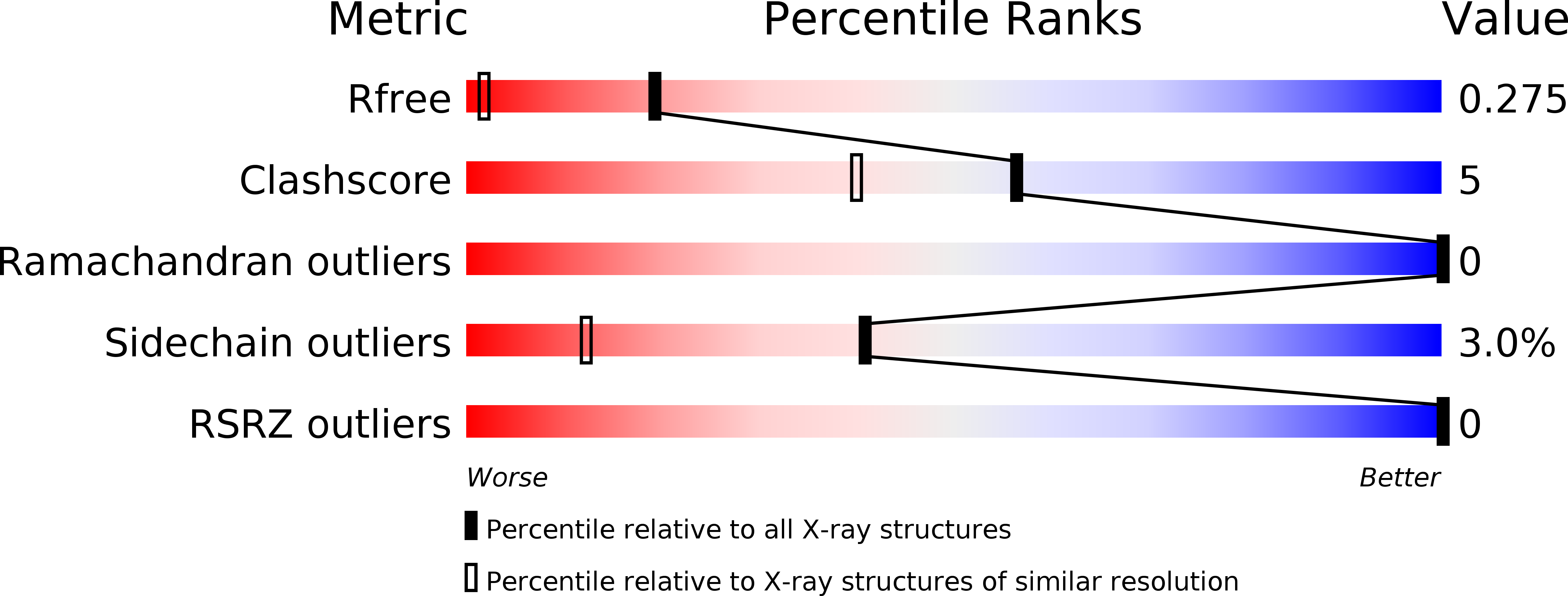
1IG5 image
Deposition Date 2001-04-17
Release Date 2001-04-25
Last Version Date 2024-02-07
Entry Detail
PDB ID:
1IG5
Title:
BOVINE CALBINDIN D9K BINDING MG2+
Biological Source:
Source Organism:
Bos taurus (Taxon ID: 9913)
Host Organism:
Method Details:
Experimental Method:
Resolution:
1.50 Å
R-Value Free:
0.28
R-Value Work:
0.19
R-Value Observed:
0.19
Space Group:
P 43 21 2
Macromolecular Entities
Polymer Type:polypeptide(L)
Molecule:VITAMIN D-DEPENDENT CALCIUM-BINDING PROTEIN, INTESTINAL
Gene (Uniprot):S100G
Chain IDs:A
Chain Length:75
Number of Molecules:1
Biological Source:Bos taurus
Ligand Molecules
Primary Citation
Structural basis for the negative allostery between Ca(2+)- and Mg(2+)-binding in the intracellular Ca(2+)-receptor calbindin D9k.
Protein Sci. 6 1139 1147 (1997)
PMID: 9194174

Abstact

The three-dimensional structures of the magnesium- and manganese-bound forms of calbindin D9k were determined to 1.6 A and 1.9 A resolution, respectively, using X-ray crystallography. These two structures are nearly identical but deviate significantly from both the calcium bound form and the metal ion-free (apo) form. The largest structural differences are seen in the C-terminal EF-hand, and involve changes in both metal ion coordination and helix packing. The N-terminal calcium binding site is not occupied by any metal ion in the magnesium and manganese structures, and shows little structural deviation from the apo and calcium bound forms. 1H-NMR and UV spectroscopic studies at physiological ion concentrations show that the C-terminal site of the protein is significantly populated by magnesium at resting cell calcium levels, and that there is a negative allosteric interaction between magnesium and calcium binding. Calcium binding was found to occur with positive cooperativity at physiological magnesium concentration.

Legend

Protein

Chemical

Disease

Primary Citation of related structures