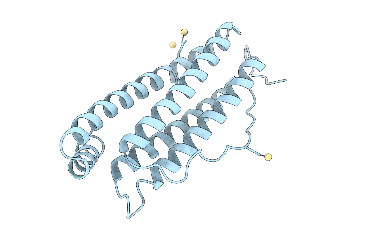
1IER image
Deposition Date 1996-05-28
Release Date 1997-01-11
Last Version Date 2024-05-22
Entry Detail
PDB ID:
1IER
Keywords:
Title:
CUBIC CRYSTAL STRUCTURE OF NATIVE HORSE SPLEEN FERRITIN
Biological Source:
Source Organism:
Equus caballus (Taxon ID: 9796)
Method Details:
Experimental Method:
Resolution:
2.26 Å
R-Value Work:
0.18
R-Value Observed:
0.18
Space Group:
F 4 3 2
Macromolecular Entities
Polymer Type:polypeptide(L)
Molecule:FERRITIN
Gene (Uniprot):FTL
Chain IDs:A
Chain Length:174
Number of Molecules:1
Biological Source:Equus caballus
Ligand Molecules
Primary Citation

Abstact

Horse-spleen apoferritin is known to crystallize in three different space groups, cubic F432, tetragonal P42(1)2 and orthorhombic P2(1)2(1)2. A structure comparison of the cubic and tetragonal forms is presented here. Both crystal forms were obtained by the vapor-diffusion technique and data were collected at 2.26 A (cubic crystal) and 2.60 A (tetragonal crystal) resolution. Two main differences were observed between these crystal structures: (i) whereas intermolecular contacts only involve salt-bridge type interactions via cadmium ions in the cubic structure, two types of interactions are observed in the tetragonal crystal (cadmium-ion-mediated salt bridges and hydrogen-bonding interactions) and (ii) cadmium ions bound in the threefold axes of ferritin molecules exhibit lower site-occupation factors in the tetragonal structure than in the cubic one.

Legend

Protein

Chemical

Disease

Primary Citation of related structures