1IE7 image
Deposition Date 2001-04-09
Release Date 2001-04-25
Last Version Date 2023-11-15
Entry Detail
PDB ID:
1IE7
Keywords:
Title:
PHOSPHATE INHIBITED BACILLUS PASTEURII UREASE CRYSTAL STRUCTURE
Biological Source:
Source Organism:
Method Details:
Experimental Method:
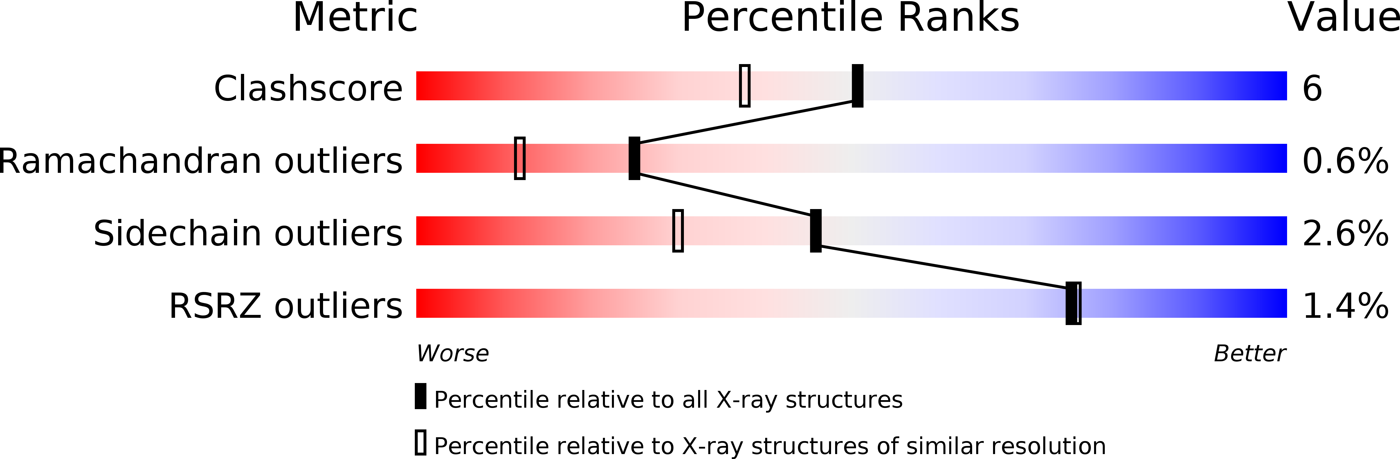
Resolution:
1.85 Å
R-Value Free:
0.21
R-Value Work:
0.17
R-Value Observed:
0.19
Space Group:
P 63 2 2
Macromolecular Entities
Polymer Type:polypeptide(L)
Molecule:UREASE GAMMA SUBUNIT
Gene (Uniprot):ureA
Chain IDs:A
Chain Length:100
Number of Molecules:1
Biological Source:Sporosarcina pasteurii
Polymer Type:polypeptide(L)
Molecule:UREASE BETA SUBUNIT
Gene (Uniprot):ureB
Chain IDs:B
Chain Length:126
Number of Molecules:1
Biological Source:Sporosarcina pasteurii
Polymer Type:polypeptide(L)
Molecule:UREASE ALPHA SUBUNIT
Gene (Uniprot):ureC
Chain IDs:C
Chain Length:570
Number of Molecules:1
Biological Source:Sporosarcina pasteurii
Modified Residue
Compound ID Chain ID Parent Comp ID Details 2D Image
CXM A MET N-CARBOXYMETHIONINE
KCX C LYS LYSINE NZ-CARBOXYLIC ACID
Primary Citation
Structure-based rationalization of urease inhibition by phosphate: novel insights into the enzyme mechanism.
J.Biol.Inorg.Chem. 6 778 790 (2001)
PMID: 11713685 DOI: 10.1007/s007750100254

Abstact

The structure of Bacillus pasteurii urease (BPU) inhibited with phosphate was solved and refined using synchrotron X-ray diffraction data from a vitrified crystal (1.85 A resolution, 99.3% completeness, data redundancy 4.6, R-factor 17.3%, PDB code 6UBP). A distance of 3.5 A separates the two Ni ions in the active site. The binding mode of the inhibitor involves the formation of four coordination bonds with the two Ni ions: one phosphate oxygen atom symmetrically bridges the two metal ions (1.9-2.0 A), while two of the remaining phosphate oxygen atoms bind to the Ni atoms at 2.4 A. The fourth phosphate oxygen is directed into the active site channel. Analysis of the H-bonding network around the bound inhibitor indicates that phosphate is bound as the H2PO4- anion, and that an additional proton is present on the Odelta2 atom of Asp(alpha363), an active site residue involved in Ni coordination through Odelta1. The flexible flap flanking the active site cavity is in the open conformation. Analysis of the complex reveals why phosphate is a relatively weak inhibitor and why sulfate does not bind to the nickels in the active site. The implications of the results for the understanding of the urease catalytic mechanism are reviewed. A novel alternative for the proton donor is presented.

Legend

Protein

Chemical

Disease

Primary Citation of related structures