
Deposition Date
2001-04-05
Release Date
2001-09-19
Last Version Date
2023-11-15
Entry Detail
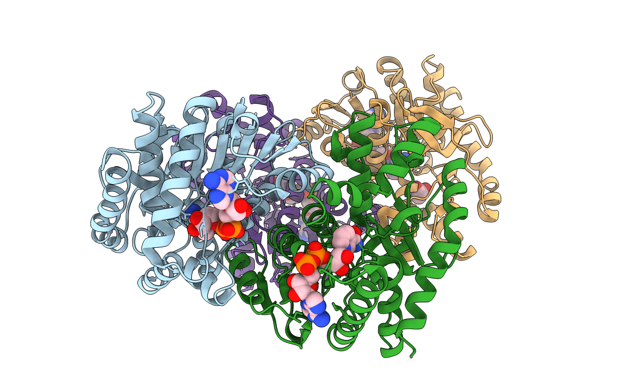
PDB ID:
1IE3
Keywords:
Title:
CRYSTAL STRUCTURE OF R153C E. COLI MALATE DEHYDROGENASE
Biological Source:
Source Organism(s):
Escherichia coli (Taxon ID: 562)
Expression System(s):
Method Details:
Experimental Method:
Resolution:
2.50 Å
R-Value Free:
0.25
R-Value Work:
0.18
R-Value Observed:
0.18
Space Group:
C 1 2 1
