1IE0 image
Deposition Date 2001-04-05
Release Date 2001-10-03
Last Version Date 2025-03-26
Entry Detail
PDB ID:
1IE0
Title:
CRYSTAL STRUCTURE OF LUXS
Biological Source:
Source Organism:
Method Details:
Experimental Method:
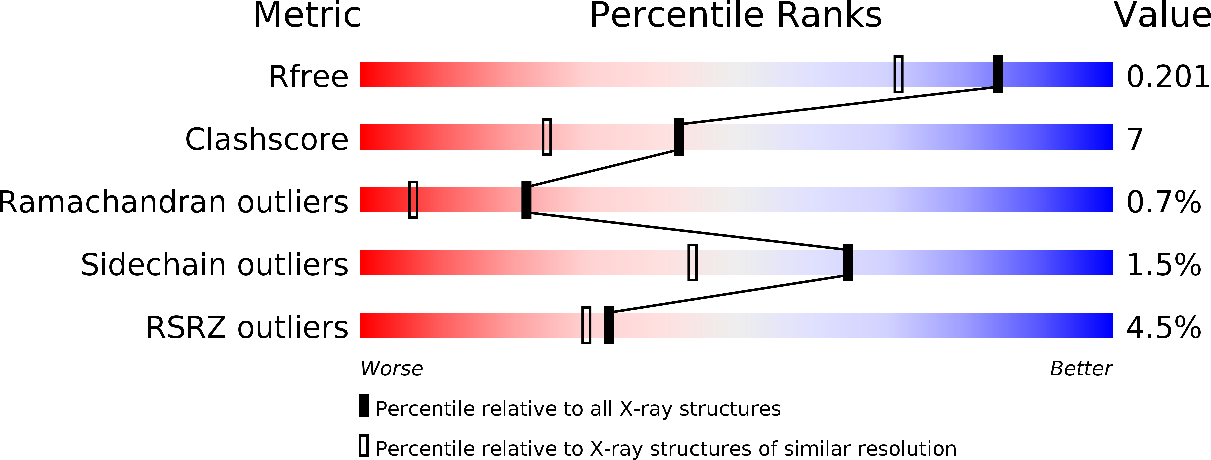
Resolution:
1.60 Å
R-Value Free:
0.20
R-Value Work:
0.17
R-Value Observed:
0.17
Space Group:
P 65 2 2
Macromolecular Entities
Structures with similar UniProt ID
Protein Blast
Polymer Type:polypeptide(L)
Molecule:AUTOINDUCER-2 PRODUCTION PROTEIN LUXS
Gene (Uniprot):luxS
Chain IDs:A
Chain Length:157
Number of Molecules:1
Biological Source:Bacillus subtilis
Modified Residue
Compound ID Chain ID Parent Comp ID Details 2D Image
OCS A CYS CYSTEINESULFONIC ACID
Primary Citation
Crystal structure of the quorum-sensing protein LuxS reveals a catalytic metal site.
Proc.Natl.Acad.Sci.USA 98 11169 11174 (2001)
PMID: 11553770 DOI: 10.1073/pnas.191223098

Abstact

The ability of bacteria to regulate gene expression in response to changes in cell density is termed quorum sensing. This behavior involves the synthesis and recognition of extracellular, hormone-like compounds known as autoinducers. Here we report the structure of an autoinducer synthase, LuxS from Bacillus subtilis, at 1.6-A resolution (R(free) = 0.204; R(work) = 0.174). LuxS is a homodimeric enzyme with a novel fold that incorporates two identical tetrahedral metal-binding sites. This metal center is composed of a Zn(2+) atom coordinated by two histidines, a cysteine, and a solvent molecule, and is reminiscent of active sites found in several peptidases and amidases. Although the nature of the autoinducer synthesized by LuxS cannot be deduced from the crystal structure, features of the putative active site suggest that LuxS might catalyze hydrolytic, but not proteolytic, cleavage of a small substrate. Our analysis represents a test of structure-based functional assignment.

Legend

Protein

Chemical

Disease

Primary Citation of related structures