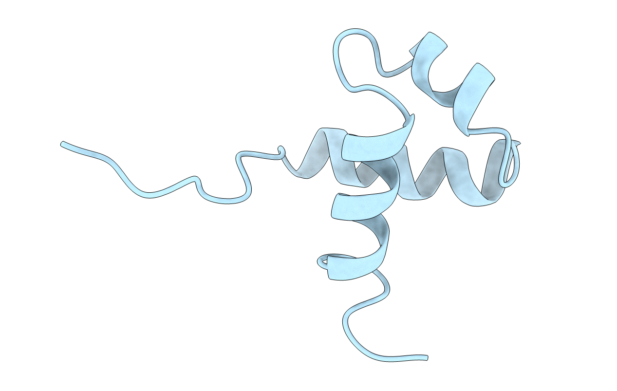
1IDY image
Deposition Date 1996-08-15
Release Date 1996-12-23
Last Version Date 2024-05-22
Entry Detail
PDB ID:
1IDY
Title:
STRUCTURE OF MYB TRANSFORMING PROTEIN, NMR, MINIMIZED AVERAGE STRUCTURE
Biological Source:
Source Organism:
Mus musculus (Taxon ID: 10090)
Host Organism:
Method Details:
Experimental Method:
Conformers Calculated:
120
Conformers Submitted:
1
Selection Criteria:
0.1 ANGSTROM MAXIMUM DISTANCE VIOLATION
Macromolecular Entities
Structures with similar UniProt ID
Protein Blast
Polymer Type:polypeptide(L)
Molecule:MOUSE C-MYB DNA-BINDING DOMAIN REPEAT 3
Gene (Uniprot):Myb
Mutagens:P140M, I155L
Chain IDs:A
Chain Length:54
Number of Molecules:1
Biological Source:Mus musculus
Ligand Molecules
Primary Citation
A small engineered protein lacks structural uniqueness by increasing the side-chain conformational entropy.
Proc.Natl.Acad.Sci.USA 93 13583 13588 (1996)
PMID: 8942977 DOI: 10.1073/pnas.93.24.13583

Abstact

A small globular protein, the third repeat of the c-Myb DNA-binding domain, which is composed of 54 amino acid residues, was engineered so as to understand the structural uniqueness of native proteins. This small protein has three alpha-helices that form a helix-turn-helix structure, which is maintained by the hydrophobic core with three Ile residues. One of the mutant proteins, with two of the buried Ile (Ile-155 and Ile-181) substituted with Leu residues, showed multiple conformations, as monitored by heteronuclear magnetic resonance spectroscopy for 13C- and 15N-labeled proteins. The increase in the side-chain conformational entropy, caused by changing the Ile to a Leu residue on an alpha-helix, could engender the lack of structural uniqueness. In native proteins, the conformations of not only the beta-branched side chains, but also those of the neighboring bulky side chains, can be greatly restricted, depending upon the local backbone structure.

Legend

Protein

Chemical

Disease

Primary Citation of related structures