Deposition Date
2001-04-03
Release Date
2001-09-05
Last Version Date
2024-11-20
Entry Detail
PDB ID:
1ID5
Keywords:
Title:
CRYSTAL STRUCTURE OF BOVINE THROMBIN COMPLEX WITH PROTEASE INHIBITOR ECOTIN
Biological Source:
Source Organism(s):
Escherichia coli (Taxon ID: 562)
Bos taurus (Taxon ID: 9913)
Bos taurus (Taxon ID: 9913)
Expression System(s):
Method Details:
Experimental Method:
Resolution:
2.50 Å
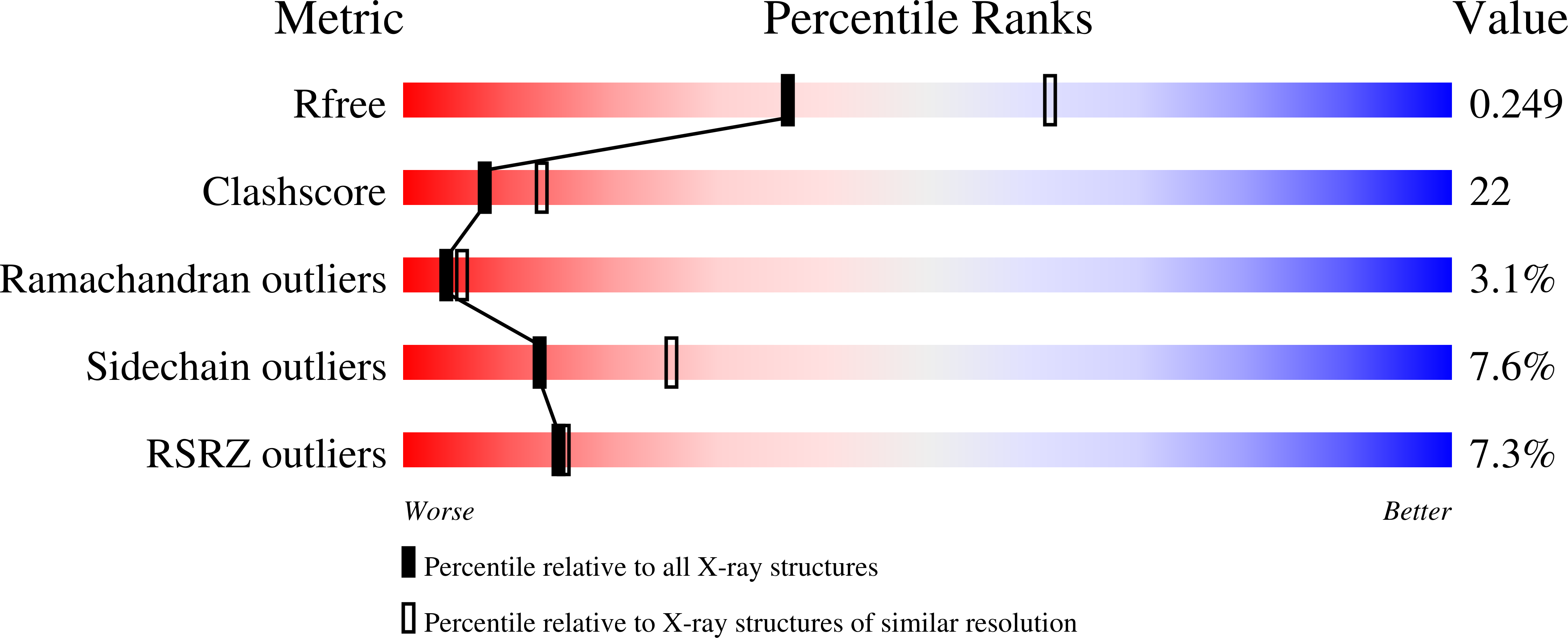
R-Value Free:
0.26
R-Value Work:
0.20
R-Value Observed:
0.20
Space Group:
C 2 2 21