
Deposition Date
2001-04-03
Release Date
2001-04-11
Last Version Date
2024-02-07
Entry Detail
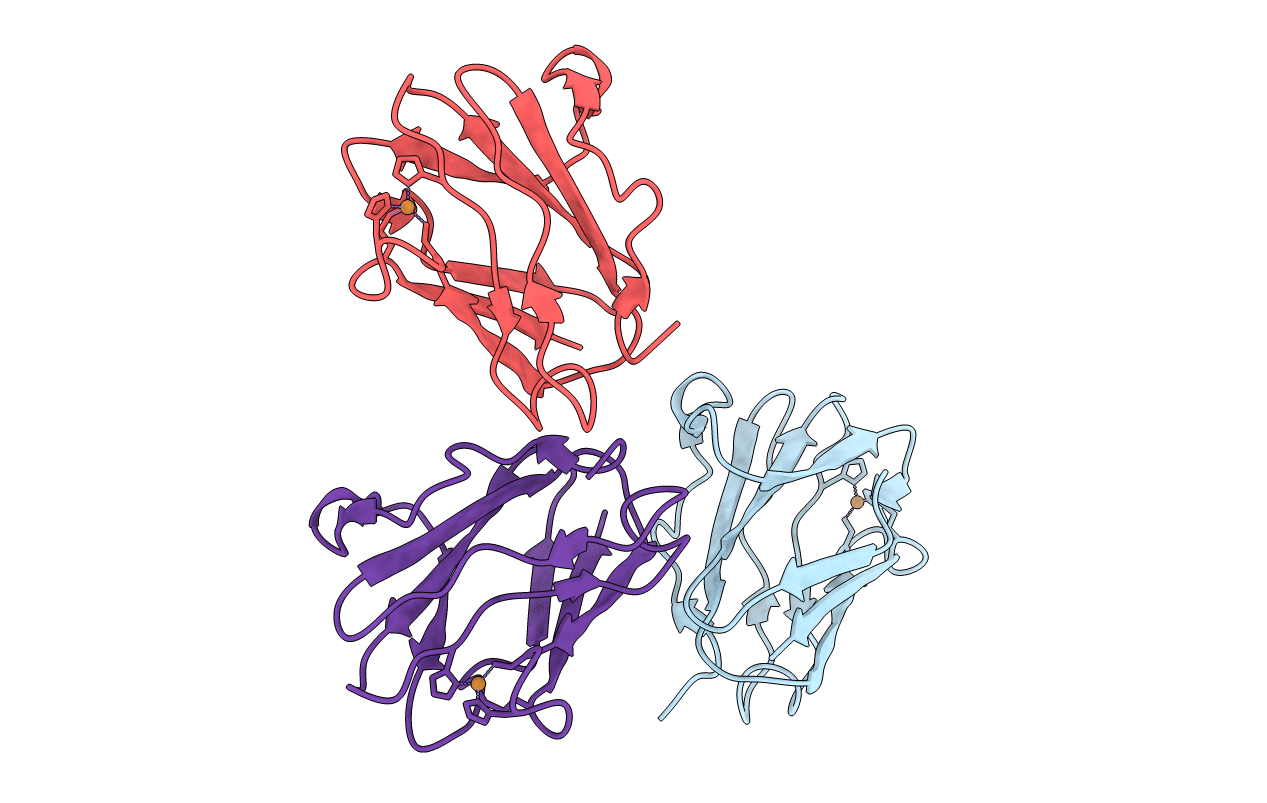
PDB ID:
1ID2
Keywords:
Title:
CRYSTAL STRUCTURE OF AMICYANIN FROM PARACOCCUS VERSUTUS (THIOBACILLUS VERSUTUS)
Biological Source:
Source Organism(s):
Paracoccus versutus (Taxon ID: 34007)
Expression System(s):
Method Details:
