Deposition Date
2001-04-02
Release Date
2001-10-17
Last Version Date
2024-02-07
Entry Detail
PDB ID:
1ID0
Keywords:
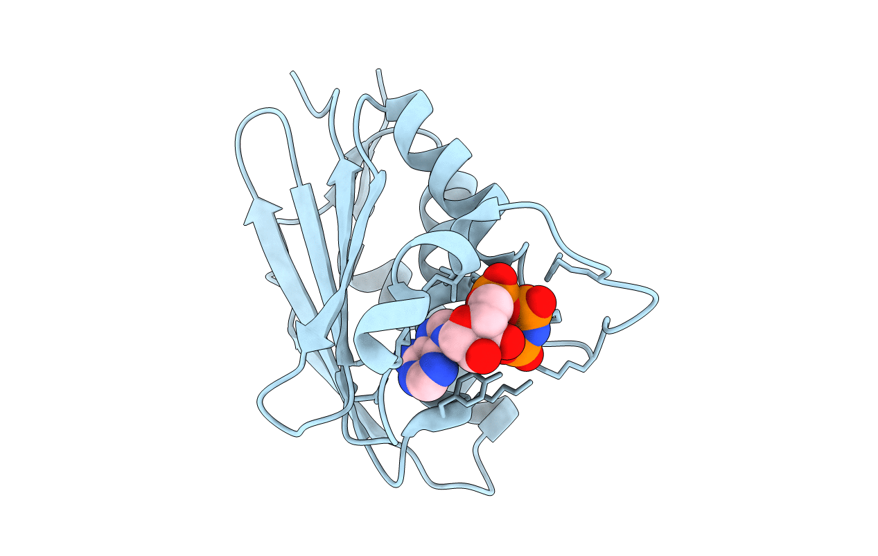
Title:
CRYSTAL STRUCTURE OF THE NUCLEOTIDE BOND CONFORMATION OF PHOQ KINASE DOMAIN
Biological Source:
Source Organism(s):
Escherichia coli (Taxon ID: 562)
Expression System(s):
Method Details:
Experimental Method:
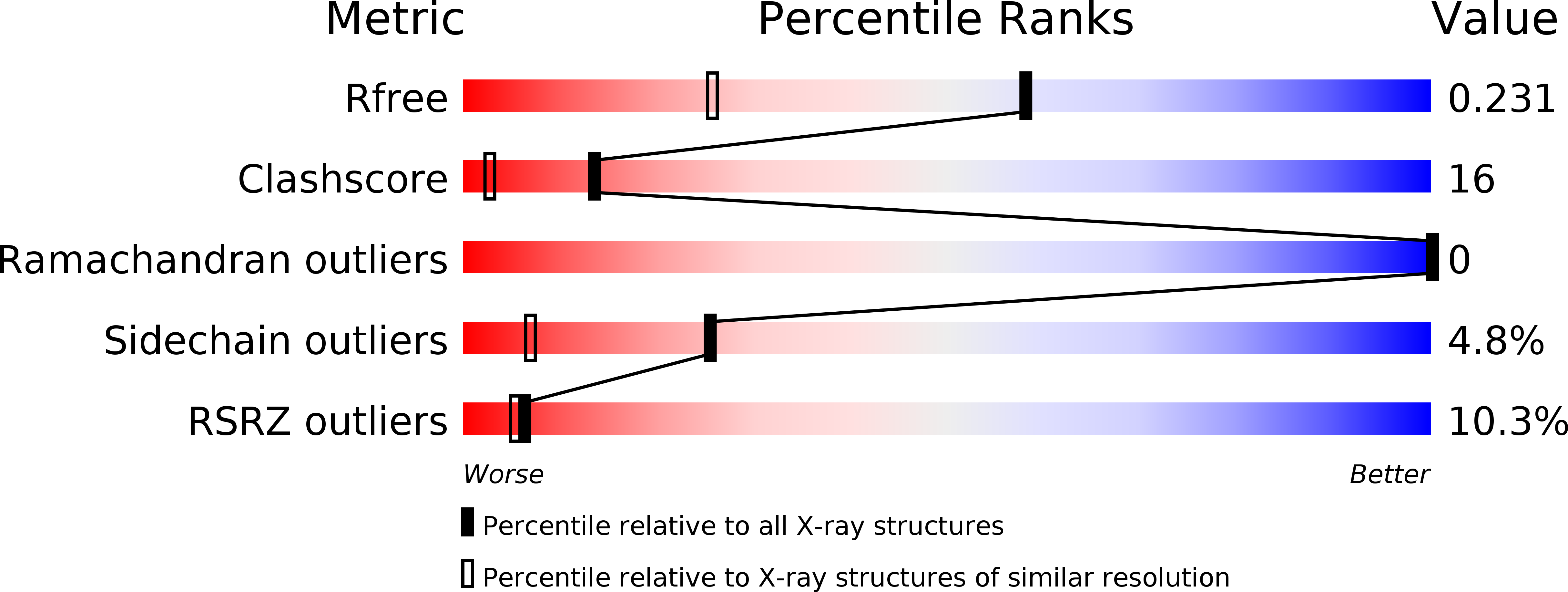
Resolution:
1.60 Å
R-Value Free:
0.23
R-Value Work:
0.19
Space Group:
P 21 21 21