
Deposition Date
2001-04-02
Release Date
2002-04-03
Last Version Date
2023-11-15
Entry Detail
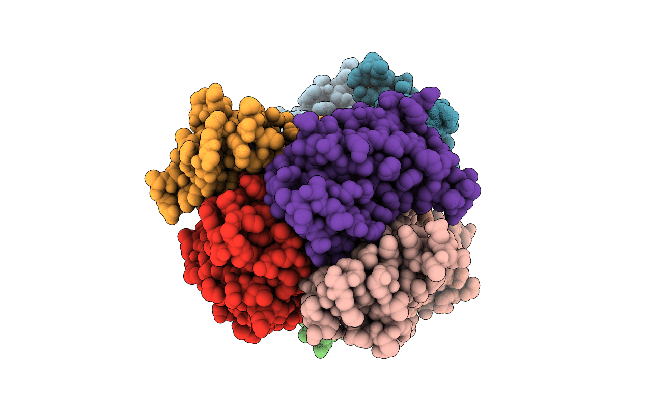
PDB ID:
1ICT
Keywords:
Title:
MONOCLINIC FORM OF HUMAN TRANSTHYRETIN COMPLEXED WITH THYROXINE (T4)
Biological Source:
Source Organism(s):
Homo sapiens (Taxon ID: 9606)
Method Details:
Experimental Method:
Resolution:
3.00 Å
R-Value Free:
0.28
R-Value Work:
0.22
R-Value Observed:
0.22
Space Group:
P 1 21 1
