Deposition Date
2001-03-29
Release Date
2001-07-25
Last Version Date
2024-02-07
Entry Detail
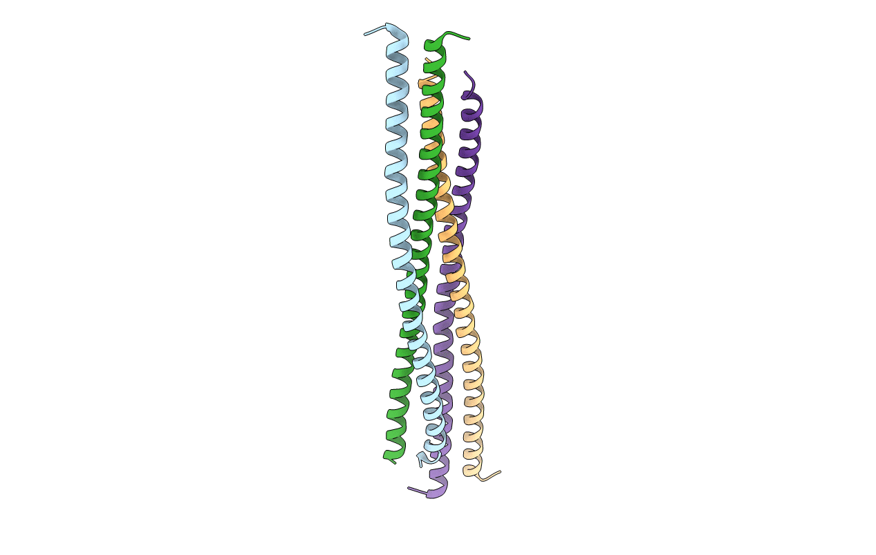
PDB ID:
1IC2
Keywords:
Title:
DECIPHERING THE DESIGN OF THE TROPOMYOSIN MOLECULE
Biological Source:
Source Organism(s):
Gallus gallus (Taxon ID: 9031)
Expression System(s):
Method Details:
Experimental Method:
Resolution:
2.00 Å
R-Value Free:
0.28
R-Value Work:
0.24
Space Group:
P 1