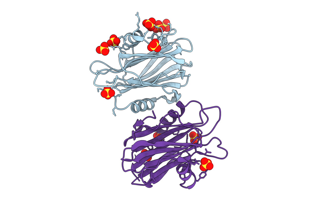
1IAZ image
Deposition Date 2001-03-24
Release Date 2001-04-04
Last Version Date 2024-02-07
Entry Detail
PDB ID:
1IAZ
Keywords:
Title:
EQUINATOXIN II
Biological Source:
Source Organism:
Actinia equina (Taxon ID: 6106)
Host Organism:
Method Details:
Experimental Method:
Resolution:
1.90 Å
R-Value Free:
0.23
R-Value Work:
0.18
R-Value Observed:
0.19
Space Group:
P 21 21 21
Macromolecular Entities
Structures with similar UniProt ID
Protein Blast
Polymer Type:polypeptide(L)
Molecule:EQUINATOXIN II
Chain IDs:A, B
Chain Length:179
Number of Molecules:2
Biological Source:Actinia equina
Ligand Molecules
Primary Citation
Crystal structure of the soluble form of equinatoxin II, a pore-forming toxin from the sea anemone Actinia equina.
Structure 9 341 346 (2001)
PMID: 11525171 DOI: 10.1016/S0969-2126(01)00592-5

Abstact

BACKGROUND: Membrane pore-forming toxins have a remarkable property: they adopt a stable soluble form structure, which, when in contact with a membrane, undergoes a series of transformations, leading to an active, membrane-bound form. In contrast to bacterial toxins, no structure of a pore-forming toxin from an eukaryotic organism has been determined so far, an indication that structural studies of equinatoxin II (EqtII) may unravel a novel mechanism. RESULTS: The crystal structure of the soluble form of EqtII from the sea anemone Actinia equina has been determined at 1.9 A resolution. EqtII is shown to be a single-domain protein based on a 12 strand beta sandwich fold with a hydrophobic core and a pair of alpha helices, each of which is associated with the face of a beta sheet. CONCLUSIONS: The structure of the 30 N-terminal residues is the largest segment that can adopt a different structure without disrupting the fold of the beta sandwich core. This segment includes a three-turn alpha helix that lies on the surface of a beta sheet and ends in a stretch of three positively charged residues, Lys-30, Arg-31, and Lys-32. On the basis of gathered data, it is suggested that this segment forms the membrane pore, whereas the beta sandwich structure remains unaltered and attaches to a membrane as do other structurally related extrinsic membrane proteins or their domains. The use of a structural data site-directed mutagenesis study should reveal the residues involved in membrane pore formation.

Legend

Protein

Chemical

Disease

Primary Citation of related structures