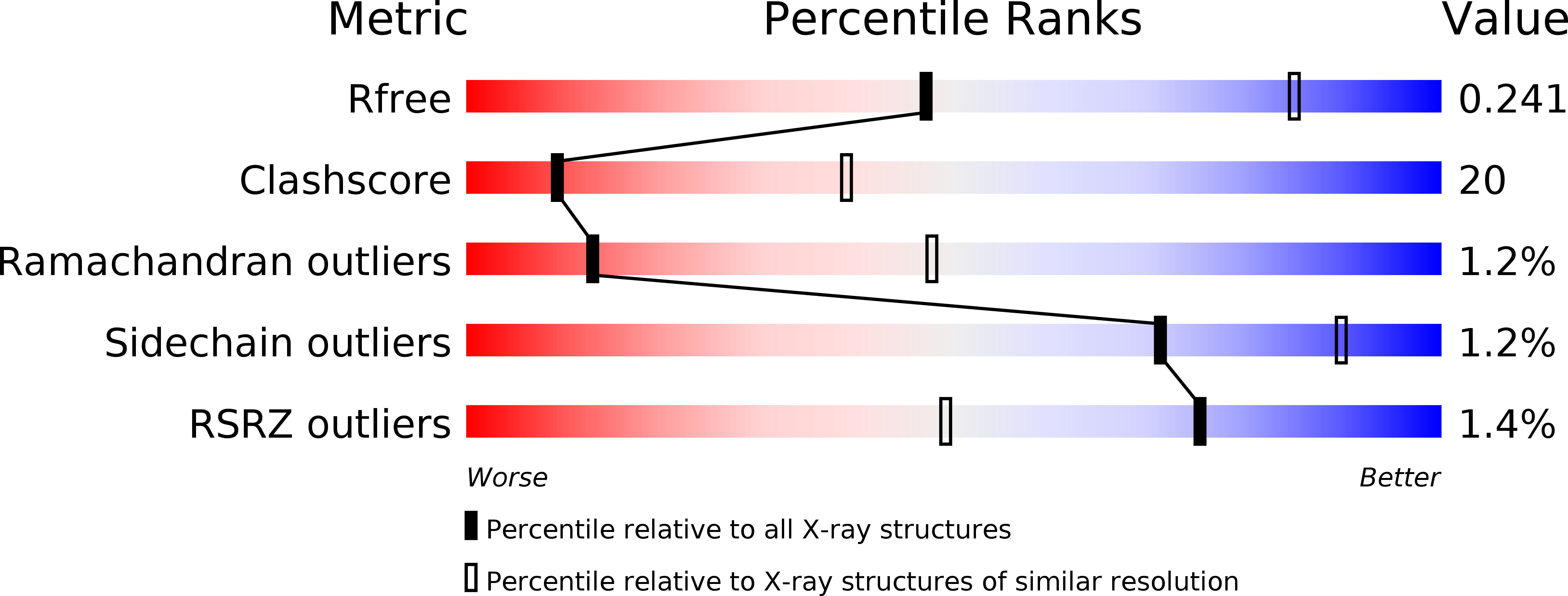
1I7X image
Deposition Date 2001-03-10
Release Date 2001-05-16
Last Version Date 2023-08-09
Entry Detail
PDB ID:
1I7X
Keywords:
Title:
BETA-CATENIN/E-CADHERIN COMPLEX
Biological Source:
Source Organism:
Mus musculus (Taxon ID: 10090)
Host Organism:
Method Details:
Experimental Method:
Resolution:
3.00 Å
R-Value Free:
0.24
R-Value Work:
0.19
R-Value Observed:
0.20
Space Group:
C 1 2 1
Macromolecular Entities
Polymer Type:polypeptide(L)
Molecule:BETA-CATENIN
Gene (Uniprot):Ctnnb1
Chain IDs:A, C
Chain Length:538
Number of Molecules:2
Biological Source:Mus musculus
Polymer Type:polypeptide(L)
Molecule:EPITHELIAL-CADHERIN
Gene (Uniprot):Cdh1
Chain IDs:B, D
Chain Length:151
Number of Molecules:2
Biological Source:Mus musculus
Primary Citation
The structure of the beta-catenin/E-cadherin complex and the molecular basis of diverse ligand recognition by beta-catenin.
Cell(Cambridge,Mass.) 105 391 402 (2001)
PMID: 11348595 DOI: 10.1016/S0092-8674(01)00330-0

Abstact

As a component of adherens junctions and the Wnt signaling pathway, beta-catenin binds cadherins, Tcf family transcription factors, and the tumor suppressor APC. We have determined the crystal structures of both unphosphorylated and phosphorylated E-cadherin cytoplasmic domain complexed with the arm repeat region of beta-catenin. The interaction spans all 12 arm repeats, and features quasi-independent binding regions that include helices which interact with both ends of the arm repeat domain and an extended stretch of 14 residues which closely resembles a portion of XTcf-3. Phosphorylation of E-cadherin results in interactions with a hydrophobic patch of beta-catenin that mimics the binding of an amphipathic XTcf-3 helix. APC contains sequences homologous to the phosphorylated region of cadherin, and is likely to bind similarly.

Legend

Protein

Chemical

Disease

Primary Citation of related structures