Deposition Date
2001-03-02
Release Date
2002-02-13
Last Version Date
2024-11-20
Entry Detail
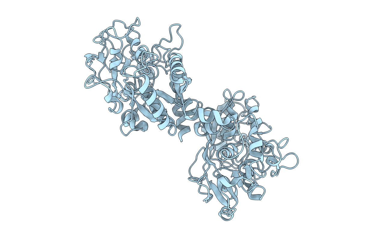
PDB ID:
1I6B
Keywords:
Title:
STRUCTURE OF EQUINE APOLACTOFERRIN AT 3.2 A RESOLUTION USING CRYSTALS GROWN AT 303K
Biological Source:
Source Organism(s):
Equus caballus (Taxon ID: 9796)
Method Details:
Experimental Method:
Resolution:
3.20 Å
R-Value Free:
0.29
R-Value Work:
0.22
R-Value Observed:
0.22
Space Group:
P 21 21 21