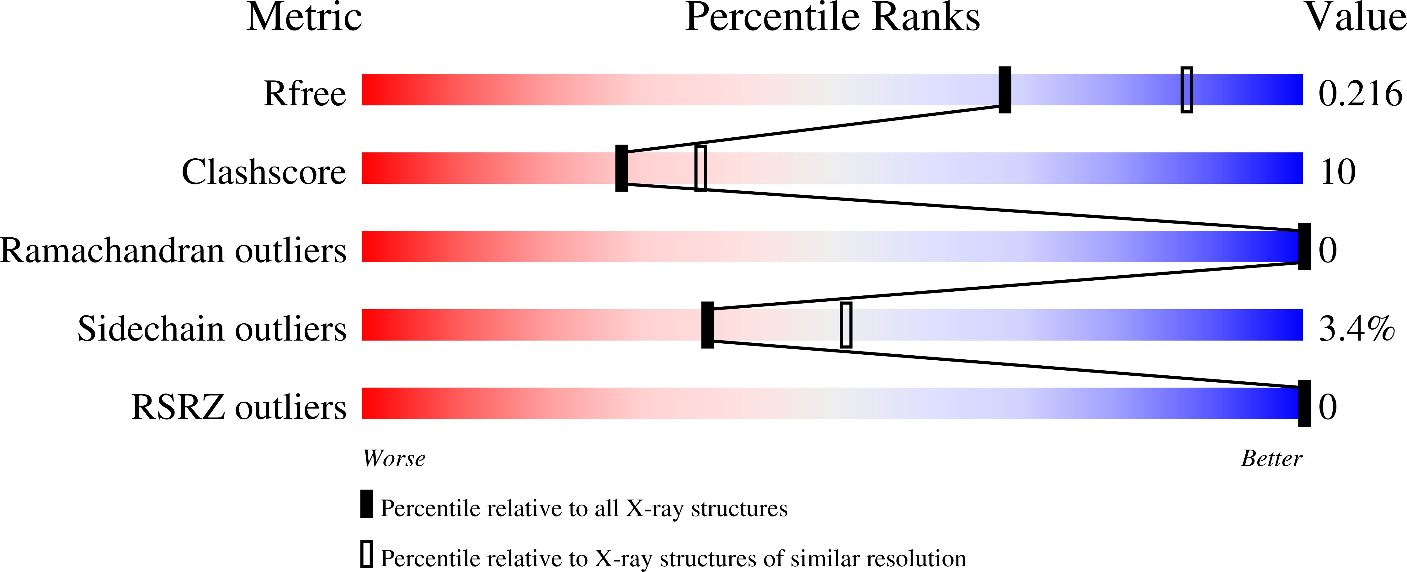
1I6A image
Deposition Date 2001-03-02
Release Date 2001-09-02
Last Version Date 2024-11-13
Entry Detail
PDB ID:
1I6A
Keywords:
Title:
CRYSTAL STRUCTURE OF THE OXIDIZED FORM OF OXYR
Biological Source:
Source Organism:
Escherichia coli (Taxon ID: 562)
Host Organism:
Method Details:
Experimental Method:
Resolution:
2.30 Å
R-Value Free:
0.21
R-Value Work:
0.16
Space Group:
C 1 2 1
Macromolecular Entities
Structures with similar UniProt ID
Protein Blast
Polymer Type:polypeptide(L)
Molecule:HYDROGEN PEROXIDE-INDUCIBLE GENES ACTIVATOR
Gene (Uniprot):oxyR
Mutagens:C143A, C180A, C259A
Chain IDs:A
Chain Length:219
Number of Molecules:1
Biological Source:Escherichia coli
Primary Citation
Structural basis of the redox switch in the OxyR transcription factor.
Cell(Cambridge,Mass.) 105 103 113 (2001)
PMID: 11301006 DOI: 10.1016/S0092-8674(01)00300-2

Abstact

The Escherichia coli OxyR transcription factor senses H2O2 and is activated through the formation of an intramolecular disulfide bond. Here we present the crystal structures of the regulatory domain of OxyR in its reduced and oxidized forms, determined at 2.7 A and 2.3 A resolutions, respectively. In the reduced form, the two redox-active cysteines are separated by approximately 17 A. Disulfide bond formation in the oxidized form results in a significant structural change in the regulatory domain. The structural remodeling, which leads to different oligomeric associations, accounts for the redox-dependent switch in OxyR and provides a novel example of protein regulation by "fold editing" through a reversible disulfide bond formation within a folded domain.

Legend

Protein

Chemical

Disease

Primary Citation of related structures