1I5S image
Deposition Date 2001-02-28
Release Date 2001-05-30
Last Version Date 2024-04-03
Entry Detail
PDB ID:
1I5S
Title:
CRYSTAL STRUCTURE OF THE KIF1A MOTOR DOMAIN COMPLEXED WITH MG-ADP
Biological Source:
Source Organism:
Mus musculus (Taxon ID: 10090)
Method Details:
Experimental Method:
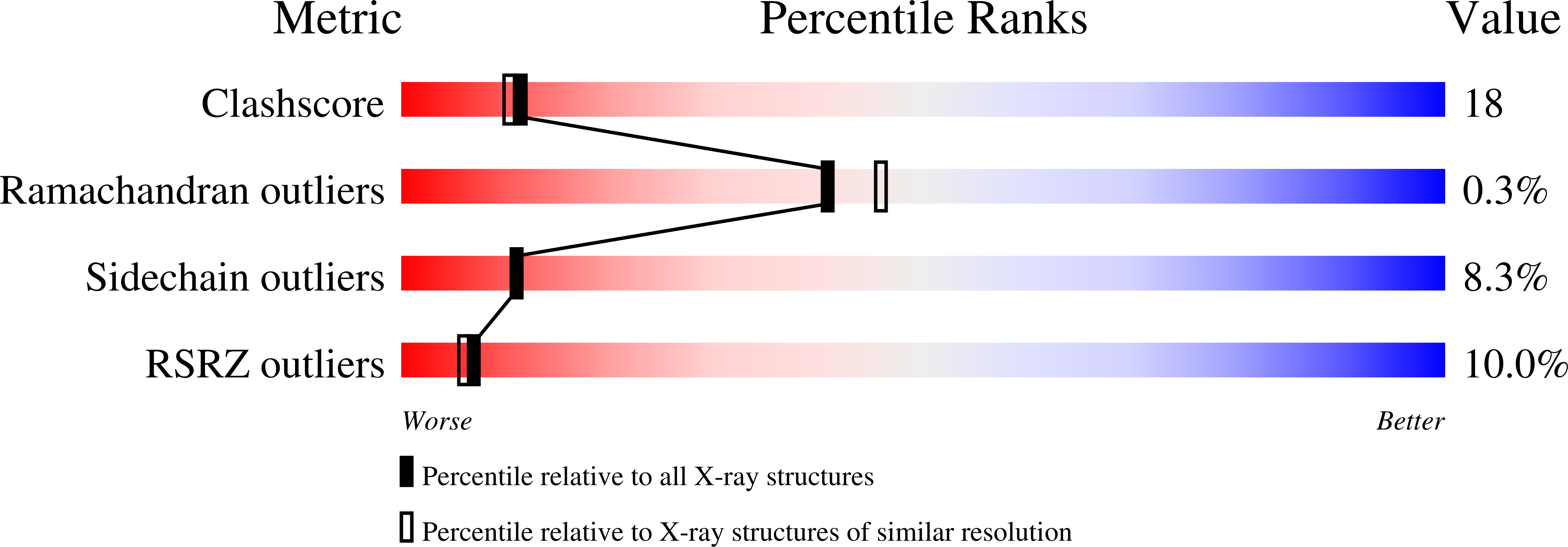
Resolution:
2.20 Å
R-Value Free:
0.22
R-Value Work:
0.21
R-Value Observed:
0.21
Space Group:
P 21 21 21
Macromolecular Entities
Polymer Type:polypeptide(L)
Molecule:KINESIN-LIKE PROTEIN KIF1A
Gene (Uniprot):Kif1a
Mutations:P202A
Chain IDs:A
Chain Length:367
Number of Molecules:1
Biological Source:Mus musculus
Primary Citation
Switch-based mechanism of kinesin motors
Nature 411 439 445 (2001)
PMID: 11373668 DOI: 10.1038/35078000

Abstact

Kinesin motors are specialized enzymes that use hydrolysis of ATP to generate force and movement along their cellular tracks, the microtubules. Although numerous biochemical and biophysical studies have accumulated much data that link microtubule-assisted ATP hydrolysis to kinesin motion, the structural view of kinesin movement remains unclear. This study of the monomeric kinesin motor KIF1A combines X-ray crystallography and cryo-electron microscopy, and allows analysis of force-generating conformational changes at atomic resolution. The motor is revealed in its two functionally critical states-complexed with ADP and with a non-hydrolysable analogue of ATP. The conformational change observed between the ADP-bound and the ATP-like structures of the KIF1A catalytic core is modular, extends to all kinesins and is similar to the conformational change used by myosin motors and G proteins. Docking of the ADP-bound and ATP-like crystallographic models of KIF1A into the corresponding cryo-electron microscopy maps suggests a rationale for the plus-end directional bias associated with the kinesin catalytic core.

Legend

Protein

Chemical

Disease

Primary Citation of related structures