Deposition Date
2001-02-22
Release Date
2001-12-19
Last Version Date
2023-08-30
Entry Detail
PDB ID:
1I4S
Keywords:
Title:
CRYSTAL STRUCTURE OF RNASE III ENDONUCLEASE DOMAIN FROM AQUIFEX AEOLICUS AT 2.15 ANGSTROM RESOLUTION
Biological Source:
Source Organism(s):
Aquifex aeolicus (Taxon ID: 63363)
Expression System(s):
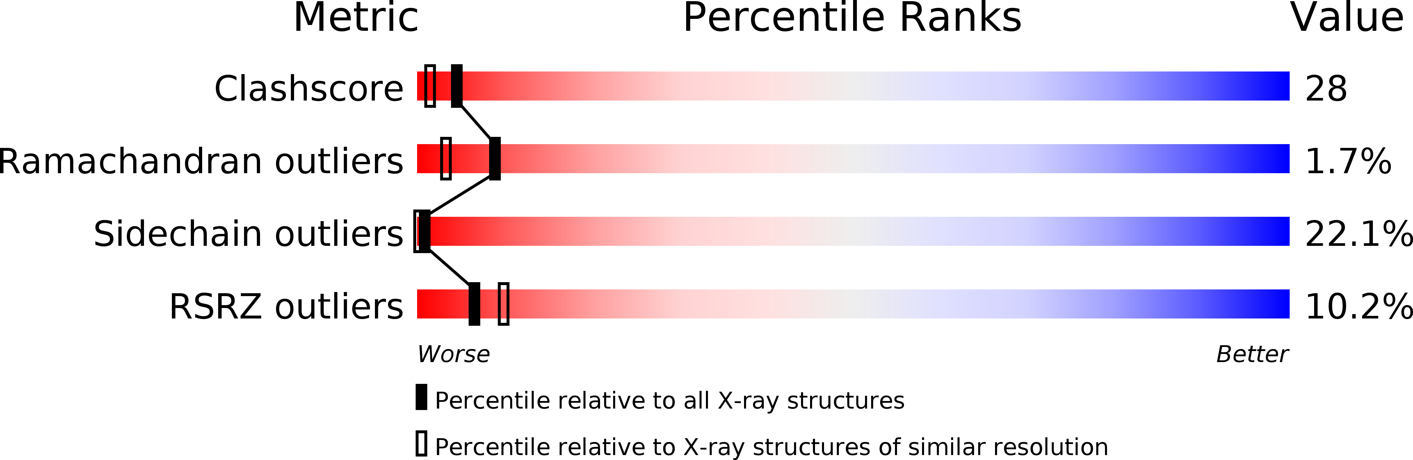
Method Details:
Experimental Method:
Resolution:
2.15 Å
R-Value Free:
0.27
R-Value Work:
0.21
R-Value Observed:
0.20
Space Group:
P 21 21 21