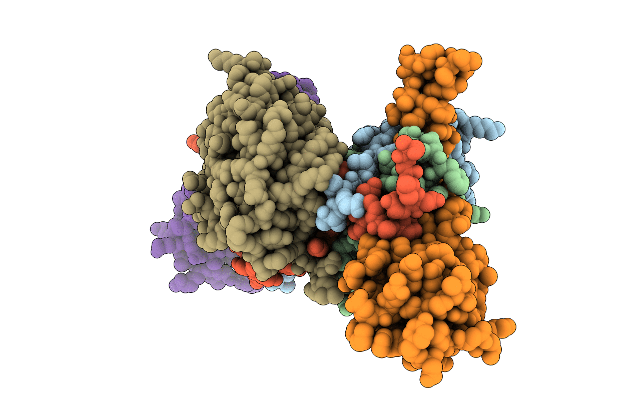
1I3O image
Entry Detail
PDB ID:
1I3O
Keywords:
Title:
CRYSTAL STRUCTURE OF THE COMPLEX OF XIAP-BIR2 AND CASPASE 3
Biological Source:
Source Organism:
PDB Version:
Deposition Date:
2001-02-15
Release Date:
2001-03-21
Method Details:
Experimental Method:
Resolution:
2.70 Å
R-Value Free:
0.27
R-Value Work:
0.24
Space Group:
P 21 21 21
Macromolecular Entities
Polymer Type:polypeptide(L)
Description:CASPASE 3
Mutations:C285A
Chain IDs:A, C
Chain Length:175
Number of Molecules:2
Biological Source:Homo sapiens
Polymer Type:polypeptide(L)
Description:CASPASE 3
Chain IDs:B, D
Chain Length:110
Number of Molecules:2
Biological Source:Homo sapiens
Polymer Type:polypeptide(L)
Description:BACULOVIRAL IAP REPEAT-CONTAINING PROTEIN 4
Mutations:C202A, C213G
Chain IDs:E, F
Chain Length:121
Number of Molecules:2
Biological Source:Homo sapiens
Ligand Molecules
Primary Citation
Structural basis for the inhibition of caspase-3 by XIAP.
Cell(Cambridge,Mass.) 104 791 800 (2001)
PMID: 11257232 DOI: 10.1016/S0092-8674(01)00274-4

Abstact

The molecular mechanism(s) that regulate apoptosis by caspase inhibition remain poorly understood. The main endogenous inhibitors are members of the IAP family and are exemplified by XIAP, which regulates the initiator caspase-9, and the executioner caspases-3 and -7. We report the crystal structure of the second BIR domain of XIAP (BIR2) in complex with caspase-3, at a resolution of 2.7 A, revealing the structural basis for inhibition. The inhibitor makes limited contacts through its BIR domain to the surface of the enzyme, and most contacts to caspase-3 originate from the N-terminal extension. This lies across the substrate binding cleft, but in reverse orientation compared to substrate binding. The mechanism of inhibition is due to a steric blockade prohibitive of substrate binding, and is distinct from the mechanism utilized by synthetic substrate analog inhibitors.

Legend

Protein

Chemical

Disease

Primary Citation of related structures