1I1C image
Deposition Date 2001-01-31
Release Date 2001-02-14
Last Version Date 2024-11-13
Entry Detail
PDB ID:
1I1C
Keywords:
Title:
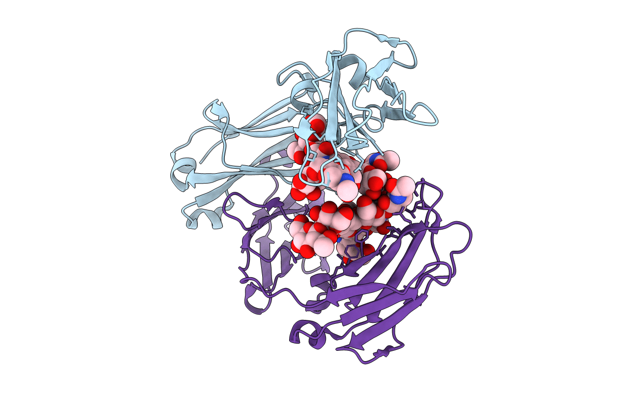
NON-FCRN BINDING FC FRAGMENT OF RAT IGG2A
Biological Source:
Source Organism:
Host Organism:
Method Details:
Experimental Method:
Resolution:
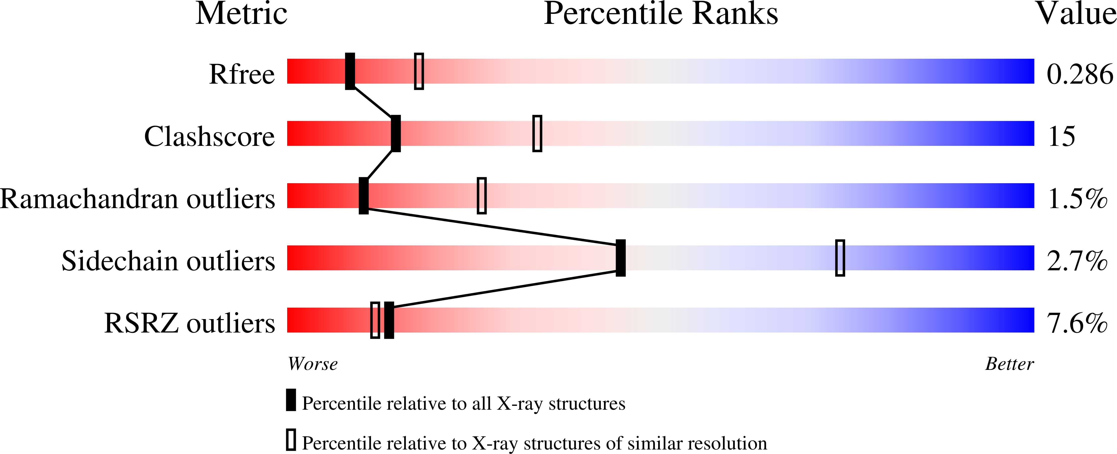
2.70 Å
R-Value Free:
0.27
R-Value Work:
0.24
Space Group:
P 1 21 1
Macromolecular Entities
Polymer Type:polypeptide(L)
Molecule:IG GAMMA-2A CHAIN C REGION
Gene (Uniprot):Igg-2a
Mutations:T252G, I253G, T254G, H310E, H433E, H435E
Chain IDs:A, B
Chain Length:239
Number of Molecules:2
Biological Source:Rattus norvegicus
Modified Residue
Compound ID Chain ID Parent Comp ID Details 2D Image
ASN B ASN GLYCOSYLATION SITE
Ligand Molecules
Primary Citation
Crystal structure at 2.8 A of an FcRn/heterodimeric Fc complex: mechanism of pH-dependent binding.
Mol.Cell 7 867 877 (2001)
PMID: 11336709 DOI: 10.1016/S1097-2765(01)00230-1

Abstact

The neonatal Fc receptor (FcRn) transports immunoglobulin G (IgG) across epithelia, binding IgG in acidic vesicles (pH < or = 6.5) and releasing IgG in the blood at pH 7.4. Well-ordered FcRn/Fc crystals are prevented by the formation of "oligomeric ribbons" of FcRn dimers bridged by Fc homodimers, thus we crystallized a 1:1 complex between rat FcRn and a heterodimeric Fc containing only one FcRn binding site. The 2.8 A complex structure demonstrates that FcRn uses its alpha2 and beta2-microglobulin domains and carbohydrate to interact with the Fc C(gamma)2-C(gamma)3 interface. The structure reveals conformational changes in Fc and three titratable salt bridges that confer pH-dependent binding, and can be used to guide rational design of therapeutic IgGs with longer serum half-lives.

Legend

Protein

Chemical

Disease

Primary Citation of related structures