1HZJ image
Deposition Date 2001-01-25
Release Date 2001-05-09
Last Version Date 2023-08-09
Entry Detail
PDB ID:
1HZJ
Keywords:
Title:
HUMAN UDP-GALACTOSE 4-EPIMERASE: ACCOMMODATION OF UDP-N-ACETYLGLUCOSAMINE WITHIN THE ACTIVE SITE
Biological Source:
Source Organism:
Homo sapiens (Taxon ID: 9606)
Host Organism:
Method Details:
Experimental Method:
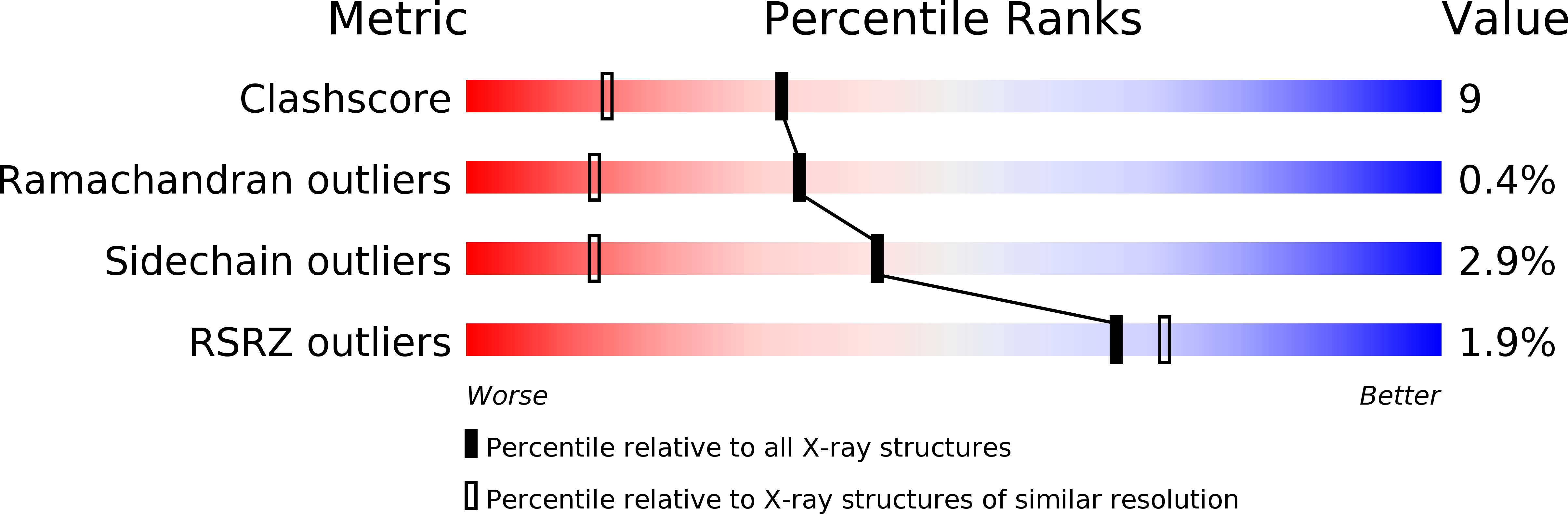
Resolution:
1.50 Å
R-Value Free:
0.22
R-Value Work:
0.18
R-Value Observed:
0.18
Space Group:
P 21 21 21
Macromolecular Entities
Structures with similar UniProt ID
Protein Blast
Polymer Type:polypeptide(L)
Molecule:UDP-GALACTOSE 4-EPIMERASE
Gene (Uniprot):GALE
Chain IDs:A, B
Chain Length:348
Number of Molecules:2
Biological Source:Homo sapiens
Primary Citation
Human UDP-galactose 4-epimerase. Accommodation of UDP-N-acetylglucosamine within the active site.
J.Biol.Chem. 276 15131 15136 (2001)
PMID: 11279032 DOI: 10.1074/jbc.M100220200

Abstact

UDP-galactose 4-epimerase catalyzes the interconversion of UDP-galactose and UDP-glucose during normal galactose metabolism. One of the key structural features in the proposed reaction mechanism for the enzyme is the rotation of a 4'-ketopyranose intermediate within the active site pocket. Recently, the three-dimensional structure of the human enzyme with bound NADH and UDP-glucose was determined. Unlike that observed for the protein isolated from Escherichia coli, the human enzyme can also turn over UDP-GlcNAc to UDP-GalNAc and vice versa. Here we describe the three-dimensional structure of human epimerase complexed with NADH and UDP-GlcNAc. To accommodate the additional N-acetyl group at the C2 position of the sugar, the side chain of Asn-207 rotates toward the interior of the protein and interacts with Glu-199. Strikingly, in the human enzyme, the structural equivalent of Tyr-299 in the E. coli protein is replaced with a cysteine residue (Cys-307) and the active site volume for the human protein is calculated to be approximately 15% larger than that observed for the bacterial epimerase. This combination of a larger active site cavity and amino acid residue replacement most likely accounts for the inability of the E. coli enzyme to interconvert UDP-GlcNAc and UDP-GalNAc.

Legend

Protein

Chemical

Disease

Primary Citation of related structures
Feedback Form
Name
Email
Institute
Feedback