1HZD image
Deposition Date 2001-01-24
Release Date 2001-12-12
Last Version Date 2023-08-09
Entry Detail
PDB ID:
1HZD
Keywords:
Title:
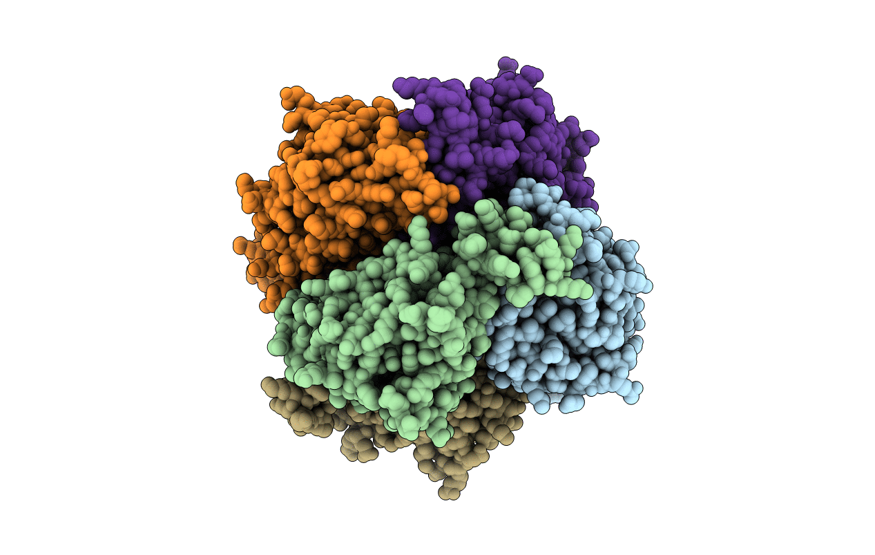
CRYSTAL STRUCTURE OF HUMAN AUH PROTEIN, AN RNA-BINDING HOMOLOGUE OF ENOYL-COA HYDRATASE
Biological Source:
Source Organism:
Homo sapiens (Taxon ID: 9606)
Method Details:
Experimental Method:
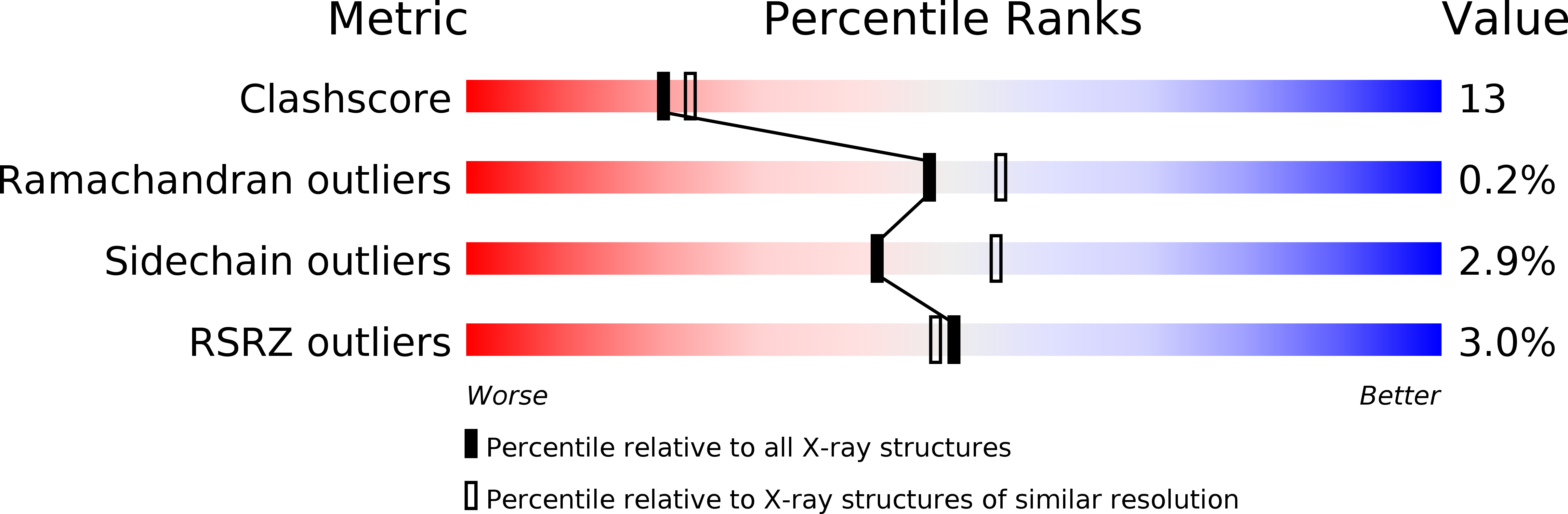
Resolution:
2.20 Å
R-Value Free:
0.25
R-Value Work:
0.20
R-Value Observed:
0.21
Space Group:
P 1 21 1
Macromolecular Entities
Structures with similar UniProt ID
Protein Blast
Polymer Type:polypeptide(L)
Molecule:AU-BINDING PROTEIN/ENOYL-COA HYDRATASE
Gene (Uniprot):AUH
Chain IDs:A, B, C, D, E, F
Chain Length:272
Number of Molecules:6
Biological Source:Homo sapiens
Primary Citation
Crystal structure of human AUH protein, a single-stranded RNA binding homolog of enoyl-CoA hydratase.
Structure 9 1253 1263 (2001)
PMID: 11738050 DOI: 10.1016/S0969-2126(01)00686-4

Abstact

BACKGROUND: The AU binding homolog of enoyl-CoA hydratase (AUH) is a bifunctional protein that has two distinct activities: AUH binds to RNA and weakly catalyzes the hydration of 2-trans-enoyl-coenzyme A (enoyl-CoA). AUH has no sequence similarity with other known RNA binding proteins, but it has considerable sequence similarity with enoyl-CoA hydratase. A segment of AUH, named the R peptide, binds to RNA. However, the mechanism of the RNA binding activity of AUH remains to be elucidated. RESULTS: We determined the crystal structure of human AUH at 2.2 A resolution. AUH adopts the typical fold of the enoyl-CoA hydratase/isomerase superfamily and forms a hexamer as a dimer of trimers. Interestingly, the surface of the AUH hexamer is positively charged, in striking contrast to the negatively charged surfaces of the other members of the superfamily. Furthermore, wide clefts are uniquely formed between the two trimers of AUH and are highly positively charged with the Lys residues in alpha helix H1, which is located on the edge of the cleft and contains the majority of the R peptide. A mutational analysis showed that the lysine residues in alpha helix H1 are essential to the RNA binding activity of AUH. CONCLUSIONS: Alpha helix H1 exposes a row of Lys residues on the solvent-accessible surface. These characteristic Lys residues are named the "lysine comb." The distances between these Lys residues are similar to those between the RNA phosphate groups, suggesting that the lysine comb may continuously bind to a single-stranded RNA. The clefts between the trimers may provide spaces sufficient to accommodate the RNA bases.

Legend

Protein

Chemical

Disease

Primary Citation of related structures