
Deposition Date
2001-01-17
Release Date
2001-04-21
Last Version Date
2024-02-07
Entry Detail
PDB ID:
1HY0
Keywords:
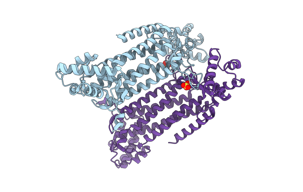
Title:
CRYSTAL STRUCTURE OF WILD TYPE DUCK DELTA 1 CRYSTALLIN (EYE LENS PROTEIN)
Biological Source:
Source Organism(s):
Anas platyrhynchos (Taxon ID: 8839)
Expression System(s):
Method Details:
Experimental Method:
Resolution:
2.20 Å
R-Value Free:
0.23
R-Value Work:
0.19
R-Value Observed:
0.20
Space Group:
P 31 2 1
