Deposition Date
1995-06-01
Release Date
1995-10-15
Last Version Date
2024-10-30
Entry Detail
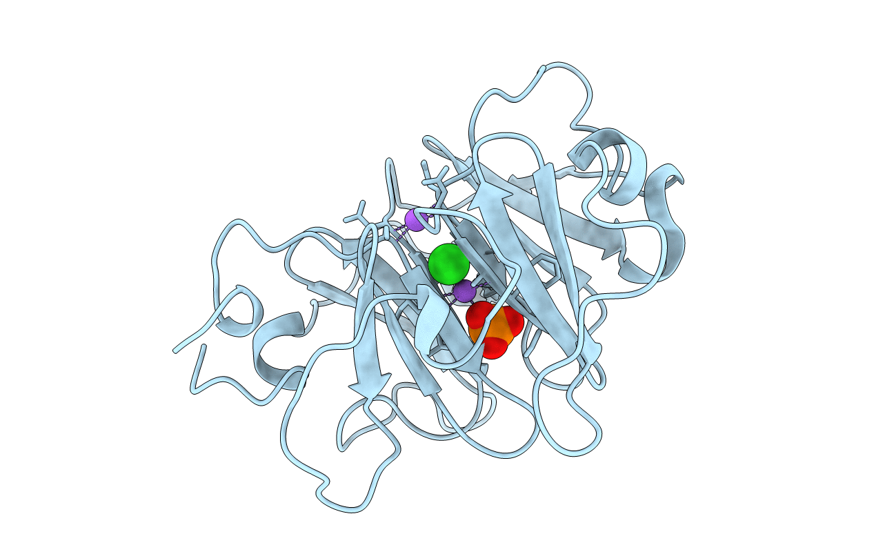
PDB ID:
1HXN
Keywords:
Title:
1.8 ANGSTROMS CRYSTAL STRUCTURE OF THE C-TERMINAL DOMAIN OF RABBIT SERUM HEMOPEXIN
Biological Source:
Source Organism:
Oryctolagus cuniculus (Taxon ID: 9986)
Method Details:
Experimental Method:
Resolution:
1.80 Å
R-Value Observed:
0.17
Space Group:
P 21 21 21