
Deposition Date
2001-01-10
Release Date
2003-06-17
Last Version Date
2023-08-09
Entry Detail
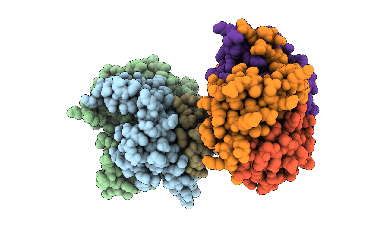
PDB ID:
1HWU
Keywords:
Title:
STRUCTURE OF PII PROTEIN FROM HERBASPIRILLUM SEROPEDICAE
Biological Source:
Source Organism(s):
Herbaspirillum seropedicae (Taxon ID: 964)
Expression System(s):
Method Details:
Experimental Method:
Resolution:
2.10 Å
R-Value Free:
0.27
R-Value Work:
0.20
Space Group:
P 21 21 21
