Deposition Date
2001-01-08
Release Date
2001-02-21
Last Version Date
2024-02-07
Entry Detail
PDB ID:
1HV9
Keywords:
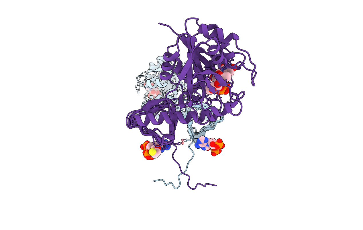
Title:
STRUCTURE OF E. COLI GLMU: ANALYSIS OF PYROPHOSPHORYLASE AND ACETYLTRANSFERASE ACTIVE SITES
Biological Source:
Source Organism(s):
Escherichia coli (Taxon ID: 562)
Expression System(s):
Method Details:
Experimental Method:
Resolution:
2.10 Å
R-Value Free:
0.24
R-Value Work:
0.21
R-Value Observed:
0.21
Space Group:
H 3 2