
Deposition Date
1993-05-27
Release Date
1994-06-22
Last Version Date
2024-10-16
Entry Detail
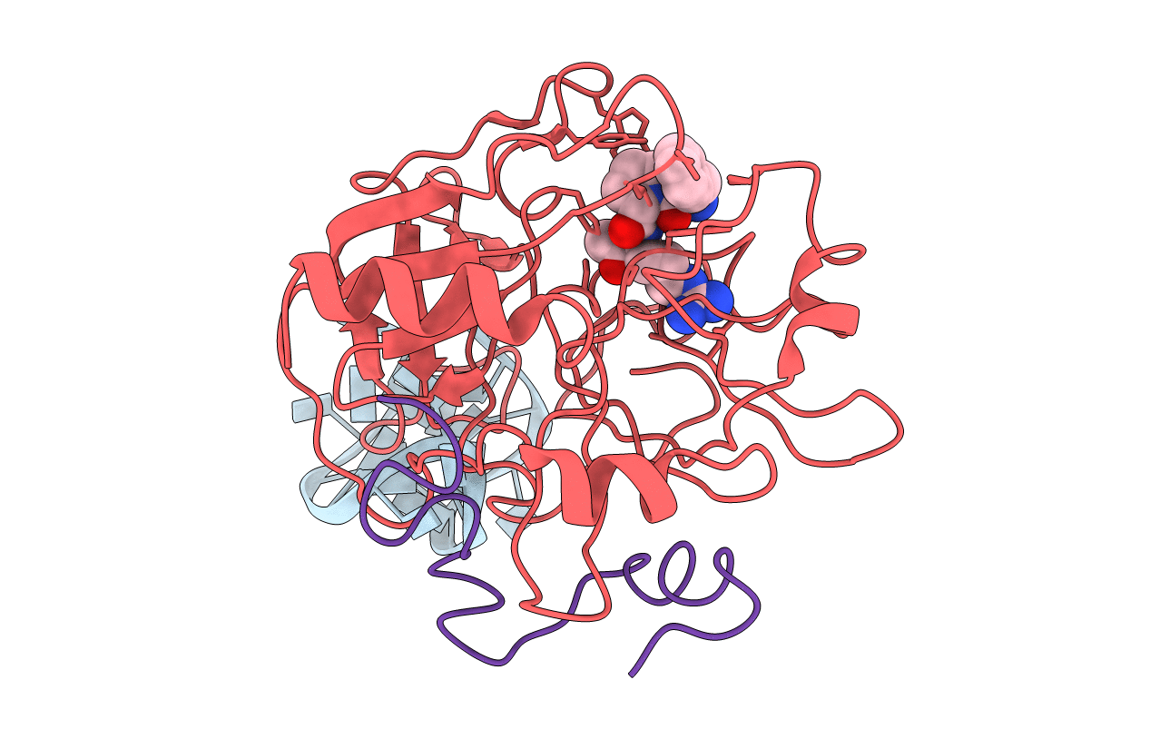
PDB ID:
1HUT
Keywords:
Title:
THE STRUCTURE OF ALPHA-THROMBIN INHIBITED BY A 15-MER SINGLE-STRANDED DNA APTAMER
Biological Source:
Source Organism(s):
Homo sapiens (Taxon ID: 9606)
Method Details:
Experimental Method:
Resolution:
2.90 Å
R-Value Work:
0.15
R-Value Observed:
0.15
Space Group:
P 21 21 21
