Deposition Date
1993-04-21
Release Date
1995-01-26
Last Version Date
2024-10-23
Entry Detail
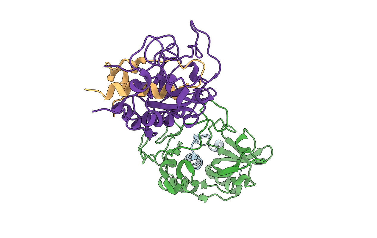
PDB ID:
1HUC
Keywords:
Title:
THE REFINED 2.15 ANGSTROMS X-RAY CRYSTAL STRUCTURE OF HUMAN LIVER CATHEPSIN B: THE STRUCTURAL BASIS FOR ITS SPECIFICITY
Biological Source:
Source Organism:
Homo sapiens (Taxon ID: 9606)
Method Details:
Experimental Method:
Resolution:
2.10 Å
R-Value Observed:
0.16
Space Group:
P 1 21 1