
Deposition Date
1996-07-10
Release Date
1997-01-11
Last Version Date
2024-11-20
Entry Detail
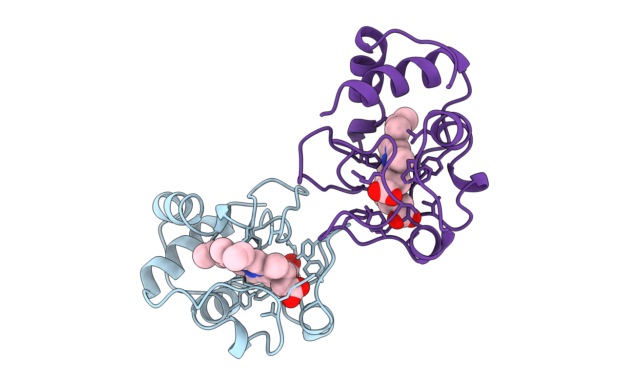
PDB ID:
1HRO
Keywords:
Title:
MOLECULAR STRUCTURE OF A HIGH POTENTIAL CYTOCHROME C2 ISOLATED FROM RHODOPILA GLOBIFORMIS
Biological Source:
Source Organism(s):
Rhodopila globiformis (Taxon ID: 1071)
Method Details:
