1HQW image
Deposition Date 2000-12-20
Release Date 2003-10-07
Last Version Date 2024-03-13
Entry Detail
PDB ID:
1HQW
Keywords:
Title:
CRYSTAL STRUCTURE OF THE COMPLEX OF CONCANAVALIN A WITH A TRIPEPTIDE YPY
Biological Source:
Source Organism:
Method Details:
Experimental Method:
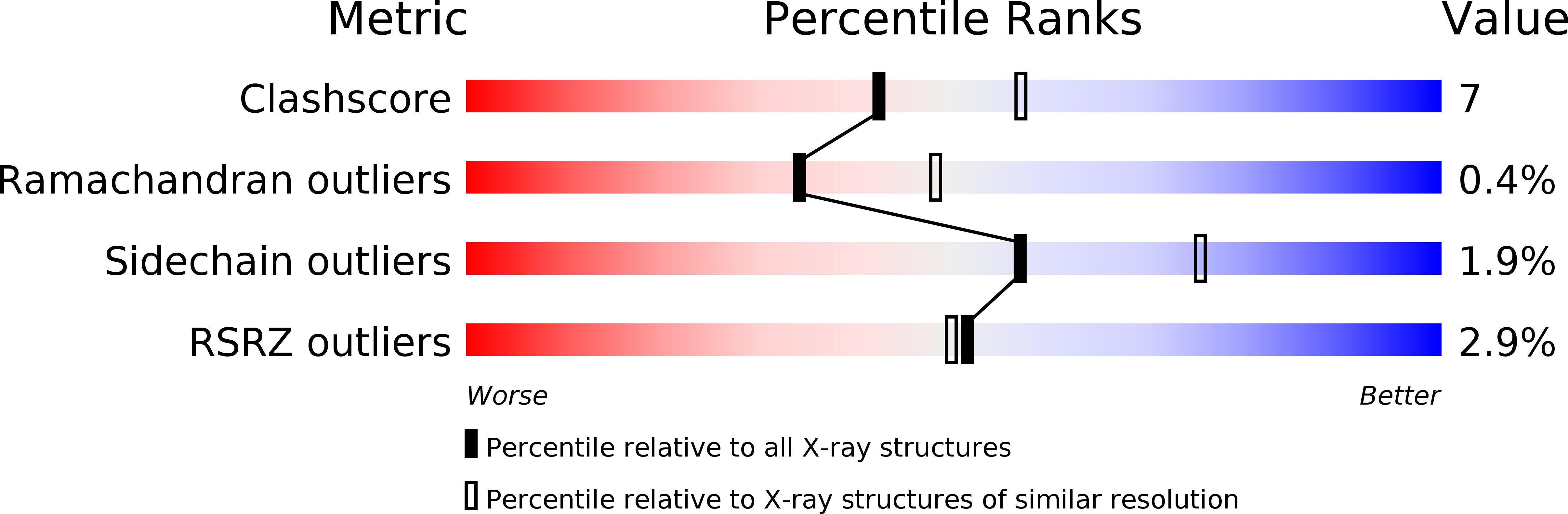
Resolution:
2.40 Å
R-Value Free:
0.23
R-Value Work:
0.17
R-Value Observed:
0.17
Space Group:
I 2 2 2
Macromolecular Entities
Structures with similar UniProt ID
Protein Blast
Polymer Type:polypeptide(L)
Molecule:CONCANAVALIN A
Chain IDs:A
Chain Length:237
Number of Molecules:1
Biological Source:Canavalia ensiformis
Polymer Type:polypeptide(L)
Molecule:YPY
Chain IDs:B
Chain Length:3
Number of Molecules:1
Biological Source:
Primary Citation
Crystal structure of the complex of concanavalin A and tripeptide
J.PROTEIN CHEM. 20 59 65 (2001)
PMID: 11330349 DOI: 10.1023/A:1011053330536

Abstact

The X-ray structure analysis of a cross-linked crystal of concanavalin A soaked with the tripeptide molecule as the probe molecule showed electron density corresponding to full occupation in the binding pocket. The site lies on the surface of concanavalin A and is surrounded by three symmetry-related molecules. The crystal structure of the tripeptide complex was refined at 2.4-A resolution to an R-factor of 17.5%, (Rfree factor of 23.7%), with an RMS deviation in bond distances of 0.01 A. The model includes all 237 residue of concanavalin A, 1 manganese ion, 1 calcium ion, 161 water molecules, 1 glutaraldehyde molecule, and 1 tripeptide molecule. This X-ray structure analysis also provides an approach to mapping the binding surface of crystalline protein with a probe molecule that is dissolved in a mixture of organic solvent with water or in neat organic solvent but is hardly dissolved in aqueous solution.

Legend

Protein

Chemical

Disease

Primary Citation of related structures