1HQL image
Deposition Date 2000-12-18
Release Date 2002-01-16
Last Version Date 2024-10-30
Entry Detail
PDB ID:
1HQL
Title:
The xenograft antigen in complex with the B4 isolectin of Griffonia simplicifolia lectin-1
Biological Source:
Source Organism:
Method Details:
Experimental Method:
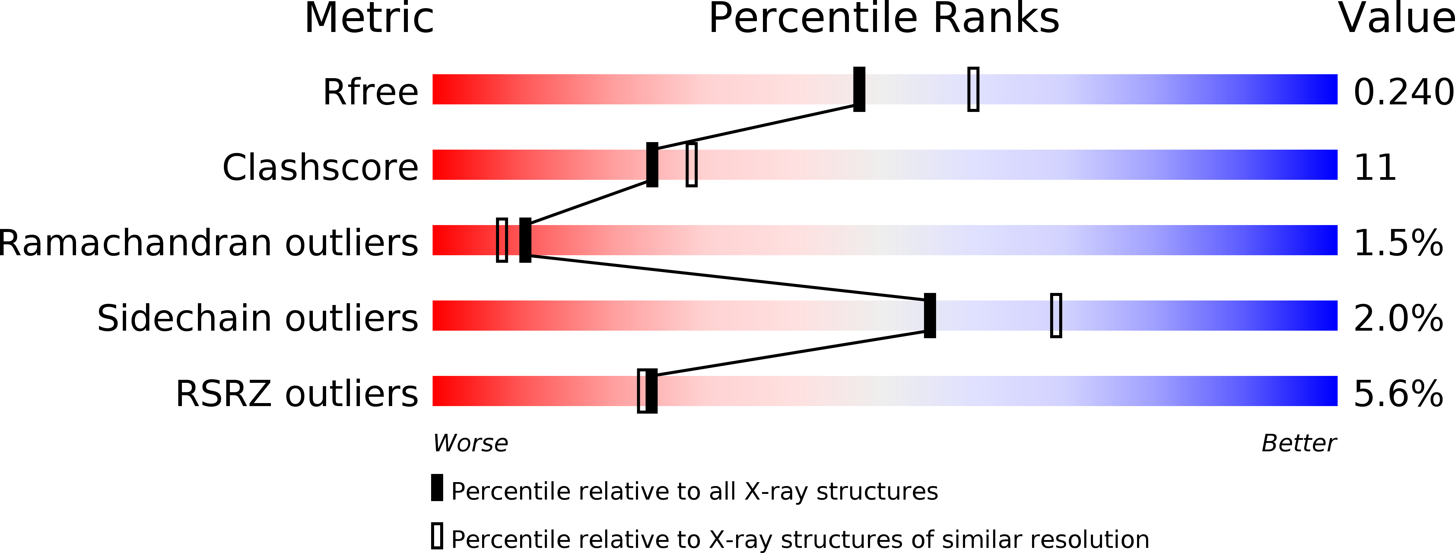
Resolution:
2.20 Å
R-Value Free:
0.25
R-Value Work:
0.23
Space Group:
P 21 21 2
Macromolecular Entities
Structures with similar UniProt ID
Protein Blast
Polymer Type:polypeptide(L)
Molecule:LECTIN
Chain IDs:A, B
Chain Length:257
Number of Molecules:2
Biological Source:Griffonia simplicifolia
Modified Residue
Compound ID Chain ID Parent Comp ID Details 2D Image
ASN A ASN GLYCOSYLATION SITE
Primary Citation
The xenograft antigen bound to Griffonia simplicifolia lectin 1-B(4). X-ray crystal structure of the complex and molecular dynamics characterization of the binding site.
J.Biol.Chem. 277 6615 6621 (2002)
PMID: 11714721 DOI: 10.1074/jbc.M109919200

Abstact

The shortage of organs for transplantation into human patients continues to be a driving force behind research into the use of tissues from non-human donors, particularly pig. The primary barrier to such xenotransplantation is the reaction between natural antibodies present in humans and Old World monkeys and the Gal alpha(1-3)Gal epitope (xenograft antigen, xenoantigen) found on the cell surfaces of the donor organ. This hyperacute immune response leads ultimately to graft rejection. Because of its high specificity for the xenograft antigen, isolectin 1-B(4) from Griffonia simplicifolia (GS-1-B(4)) has been used as an immunodiagnostic reagent. Furthermore, haptens that inhibit natural antibodies also inhibit GS-1-B(4) from binding to the xenoantigen. Here we report the first x-ray crystal structure of the xenograft antigen bound to a protein (GS-1-B(4)). The three-dimensional structure was determined from orthorhombic crystals at a resolution of 2.3 A. To probe the influence of binding on ligand properties, we report also the results of molecular dynamics (MD) simulations on this complex as well as on the free ligand. The MD simulations were performed with the AMBER force-field for proteins augmented with the GLYCAM parameters for glycosides and glycoproteins. The simulations were performed for up to 10 ns in the presence of explicit solvent. Through comparison with MD simulations performed for the free ligand, it has been determined that GS-1-B(4) recognizes the lowest energy conformation of the disaccharide. In addition, the x-ray and modeling data provide clear explanations for the reported specificities of the GS-1-B(4) lectin. It is anticipated that a further understanding of the interactions involving the xenograft antigen will help in the development of therapeutic agents for application in the prevention of hyperacute xenograft rejection.

Legend

Protein

Chemical

Disease

Primary Citation of related structures