Deposition Date
2000-12-08
Release Date
2001-03-07
Last Version Date
2024-10-30
Entry Detail
PDB ID:
1HO3
Keywords:
Title:
CRYSTAL STRUCTURE ANALYSIS OF E. COLI L-ASPARAGINASE II (Y25F MUTANT)
Biological Source:
Source Organism(s):
Escherichia coli (Taxon ID: 562)
Expression System(s):
Method Details:
Experimental Method:
Resolution:
2.50 Å
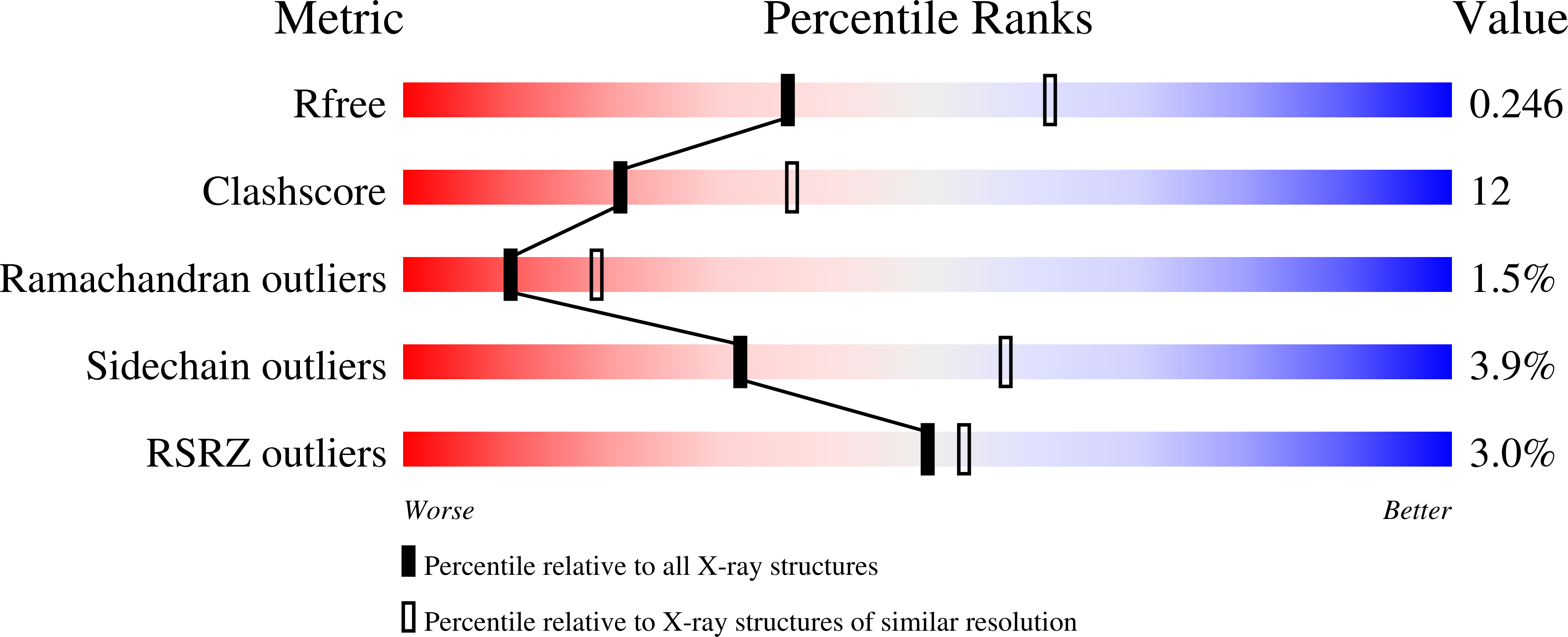
R-Value Free:
0.24
R-Value Work:
0.18
Space Group:
P 65 2 2