1HNV image
Deposition Date 1995-03-30
Release Date 1995-07-10
Last Version Date 2024-02-07
Entry Detail
PDB ID:
1HNV
Title:
STRUCTURE OF HIV-1 RT(SLASH)TIBO R 86183 COMPLEX REVEALS SIMILARITY IN THE BINDING OF DIVERSE NONNUCLEOSIDE INHIBITORS
Biological Source:
Source Organism:
Host Organism:
Method Details:
Experimental Method:
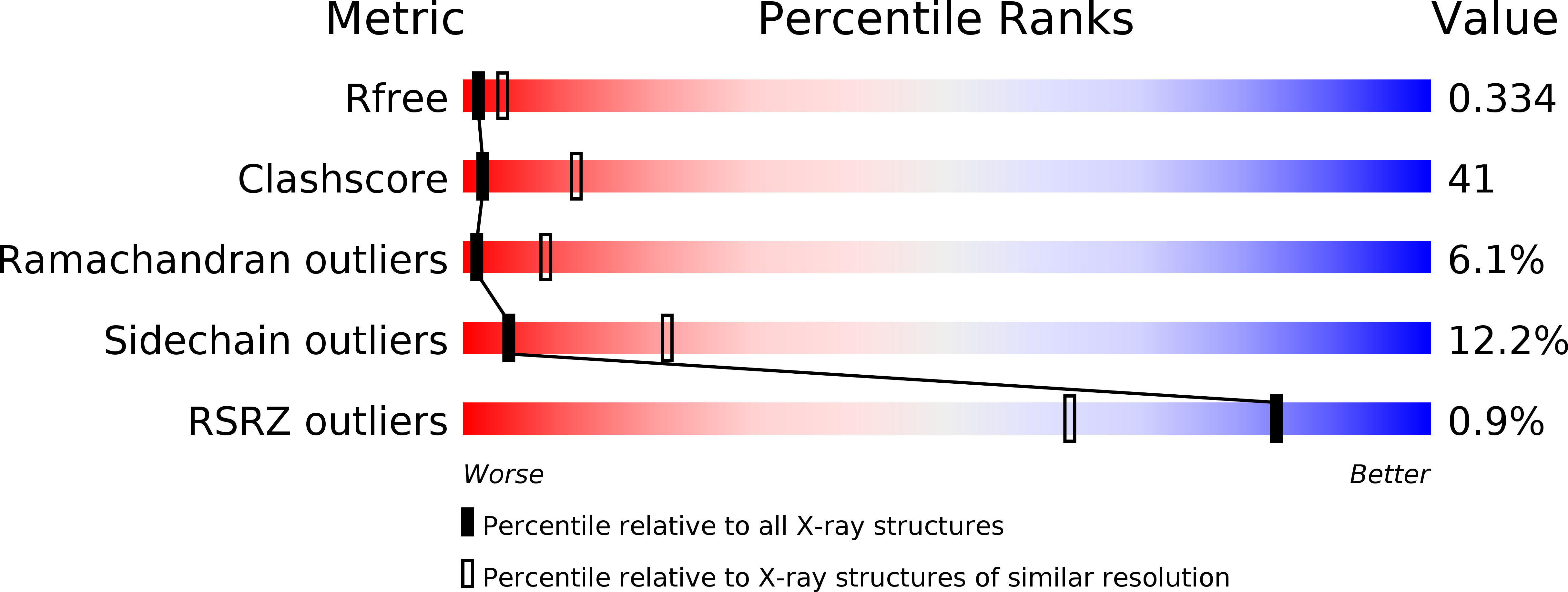
Resolution:
3.00 Å
R-Value Free:
0.35
R-Value Work:
0.24
R-Value Observed:
0.24
Space Group:
C 1 2 1
Macromolecular Entities
Polymer Type:polypeptide(L)
Molecule:HIV-1 REVERSE TRANSCRIPTASE (SUBUNIT P66)
Gene (Uniprot):gag-pol
Chain IDs:A
Chain Length:558
Number of Molecules:1
Biological Source:Human immunodeficiency virus 1
Polymer Type:polypeptide(L)
Molecule:HIV-1 REVERSE TRANSCRIPTASE (SUBUNIT P51)
Gene (Uniprot):gag-pol
Chain IDs:B
Chain Length:427
Number of Molecules:1
Biological Source:Human immunodeficiency virus 1
Ligand Molecules
Primary Citation

Abstact

We report the structure of HIV-1 reverse transcriptase (RT) complexed with the nonnucleoside inhibitor TIBO R 86183 at 3.0 A resolution. Comparing this structure with those of complexes of HIV-1 RT/alpha-APA R 95845 and HIV-1 RT/nevirapine provides a basis for understanding the nature of nonnucleoside inhibitor binding, the structure of the binding site and the interactions between the bound inhibitors and surrounding amino acid residues as well as for understanding mechanisms of inhibition by and resistance to nonnucleoside inhibitors. All three inhibitors considered assume a similar butterfly-like shape and bind to HIV-1 RT in a very similar way. Important differences occur in the conformation of amino acid residues that form the binding pocket.

Legend

Protein

Chemical

Disease

Primary Citation of related structures