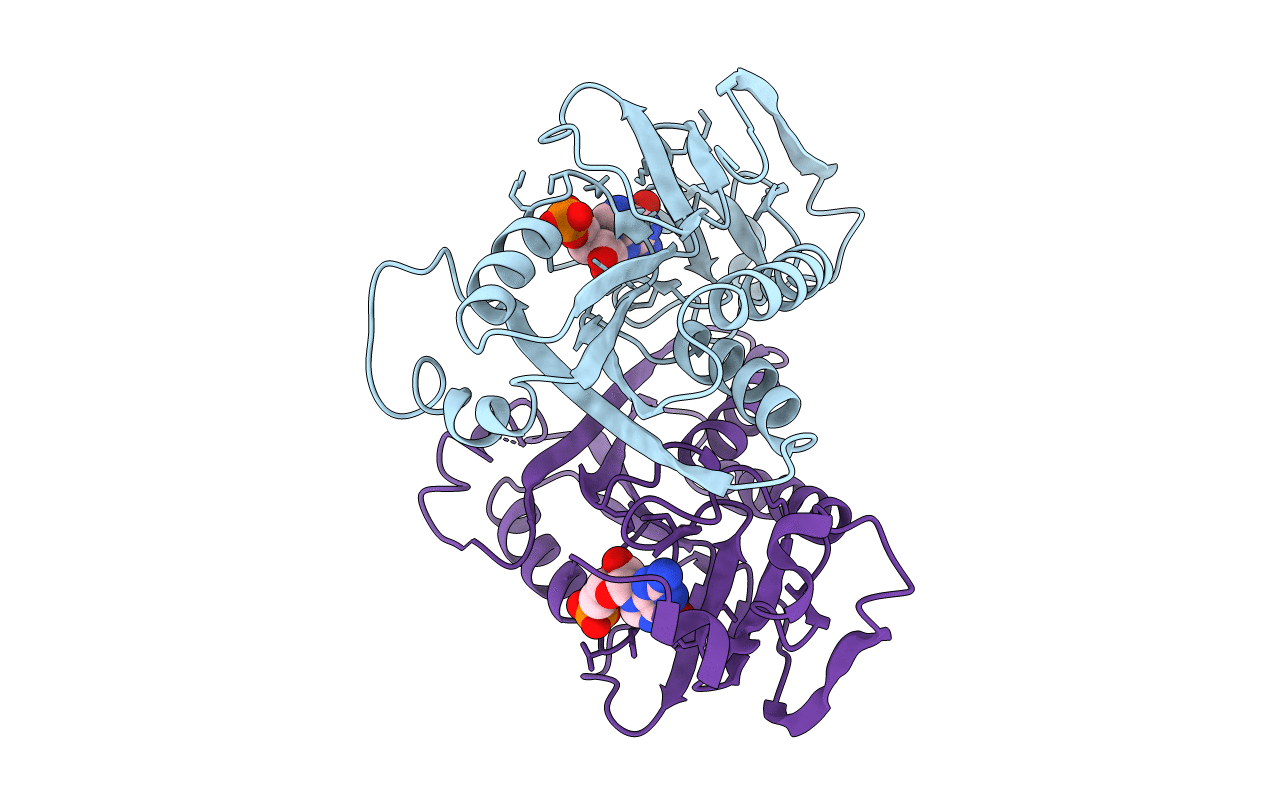
1HMP image
Deposition Date 1994-06-03
Release Date 1995-06-03
Last Version Date 2024-02-07
Entry Detail
PDB ID:
1HMP
Title:
THE CRYSTAL STRUCTURE OF HUMAN HYPOXANTHINE-GUANINE PHOSPHORIBOSYLTRANSFERASE WITH BOUND GMP
Biological Source:
Source Organism:
Homo sapiens (Taxon ID: 9606)
Method Details:
Experimental Method:
Resolution:
2.50 Å
R-Value Work:
0.18
R-Value Observed:
0.18
Space Group:
P 21 21 2
Macromolecular Entities
Polymer Type:polypeptide(L)
Molecule:HYPOXANTHINE GUANINE PHOSPHORIBOSYL-TRANSFERASE
Gene (Uniprot):HPRT1
Chain IDs:A, B
Chain Length:217
Number of Molecules:2
Biological Source:Homo sapiens
Ligand Molecules
Primary Citation
The crystal structure of human hypoxanthine-guanine phosphoribosyltransferase with bound GMP.
Cell(Cambridge,Mass.) 78 325 334 (1994)
PMID: 8044844 DOI: 10.1016/0092-8674(94)90301-8

Abstact

The crystal structure of HGPRTase with bound GMP has been determined and refined to 2.5 A resolution. The enzyme has a core alpha/beta structure resembling the nucleotide-binding fold of dehydrogenases, and a second lobe composed of residues from the amino and carboxy termini. The GMP molecule binds in an anti conformation in a solvent-exposed cleft of the enzyme. Lys-165, which forms a hydrogen bond to O6 of GMP, appears to be critical for determining the specificity for guanine and hypoxanthine over adenine. The location of active site residues also provides evidence for a possible mechanism for general base-assisted HGPRTase catalysis. A rationalization of the effects on stability and activity of naturally occurring single amino acid mutations of HGPRTase is presented, including a discussion of several mutations at the active site that lead to Lesch-Nyhan syndrome.

Legend

Protein

Chemical

Disease

Primary Citation of related structures