Deposition Date
1993-10-13
Release Date
1994-04-30
Last Version Date
2024-02-07
Entry Detail
PDB ID:
1HLC
Keywords:
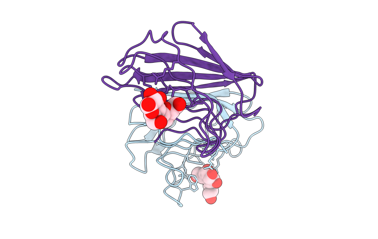
Title:
X-RAY CRYSTAL STRUCTURE OF THE HUMAN DIMERIC S-LAC LECTIN, L-14-II, IN COMPLEX WITH LACTOSE AT 2.9 ANGSTROMS RESOLUTION
Biological Source:
Source Organism(s):
Homo sapiens (Taxon ID: 9606)
Method Details:
Experimental Method:
Resolution:
2.90 Å
R-Value Work:
0.17
R-Value Observed:
0.17
Space Group:
P 21 21 21