Deposition Date
1994-03-22
Release Date
1994-06-22
Last Version Date
2024-11-13
Entry Detail
PDB ID:
1HLB
Keywords:
Title:
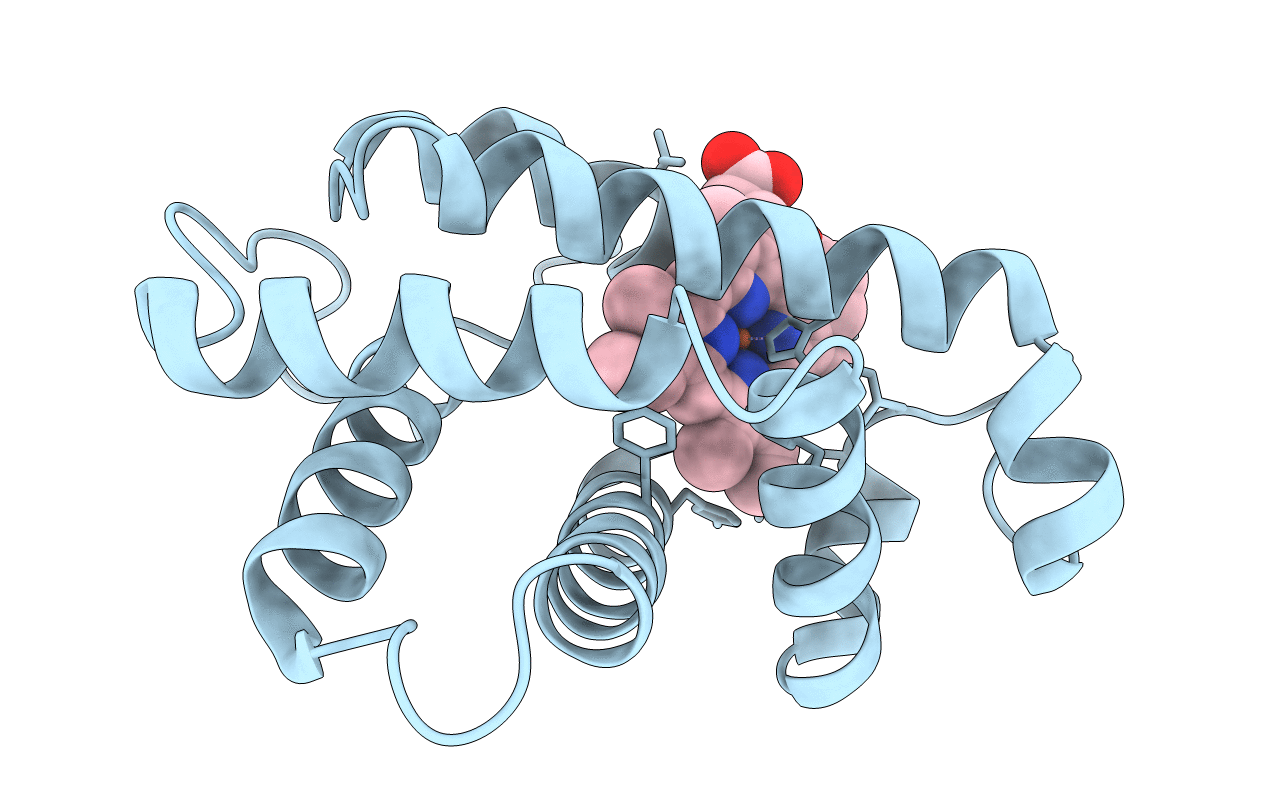
Structural analysis of monomeric hemichrome and dimeric cyanomet hemoglobins from Caudina arenicola
Biological Source:
Source Organism(s):
Caudina arenicola (Taxon ID: 7698)
Method Details:
Experimental Method:
Resolution:
2.50 Å
R-Value Work:
0.15
R-Value Observed:
0.15
Space Group:
P 1 21 1