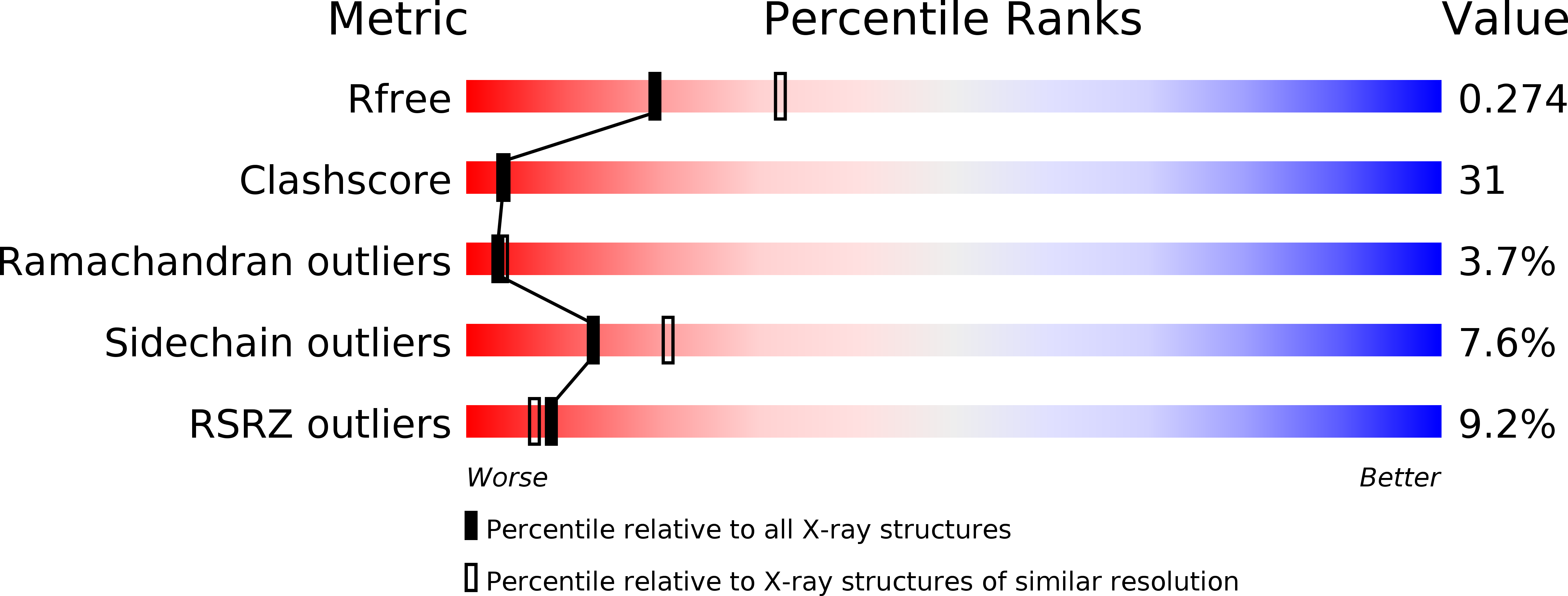
1HKX image
Deposition Date 2003-03-12
Release Date 2003-06-02
Last Version Date 2024-06-19
Entry Detail
PDB ID:
1HKX
Keywords:
Title:
Crystal structure of calcium/calmodulin-dependent protein kinase
Biological Source:
Source Organism:
MUS MUSCULUS (Taxon ID: 10090)
Host Organism:
Method Details:
Experimental Method:
Resolution:
2.65 Å
R-Value Free:
0.27
R-Value Work:
0.24
R-Value Observed:
0.24
Space Group:
C 1 2 1
Macromolecular Entities
Structures with similar UniProt ID
Protein Blast
Polymer Type:polypeptide(L)
Molecule:CALCIUM/CALMODULIN-DEPENDENT PROTEIN KINASE TYPE II ALPHA CHAIN
Gene (Uniprot):Camk2a
Chain IDs:A, B, C, D, E, F, G, H, I, J, K, L, M, N
Chain Length:147
Number of Molecules:14
Biological Source:MUS MUSCULUS
Primary Citation
Crystal Structure of a Tetradecameric Assembly of the Association Domain of Ca2+/Calmodulin-Dependent Kinase II
Mol.Cell 11 1241 ? (2003)
PMID: 12769848 DOI: 10.1016/S1097-2765(03)00171-0

Abstact

We report the crystal structure of the 143 residue association domain of Ca(2+)/calmodulin-dependent protein kinase II (CaMKII). The association domain forms a hub-like assembly, composed of two rings of seven protomers each, which are stacked head to head and held together by extensive interfaces. The tetradecameric organization of the assembly was confirmed by analytical ultracentrifugation and multiangle light scattering. Individual protomers form wedge-shaped structures from which N-terminal helical segments that connect to the kinase domain extend toward the equatorial plane of the assembly, consistent with the arrangement of the kinase domains in a second outer ring. A deep and highly conserved pocket present within the association domain may serve as a docking site for proteins that interact with CaMKII.

Legend

Protein

Chemical

Disease

Primary Citation of related structures