1HJR image
Deposition Date 1994-12-02
Release Date 1995-02-07
Last Version Date 2024-02-07
Entry Detail
PDB ID:
1HJR
Title:
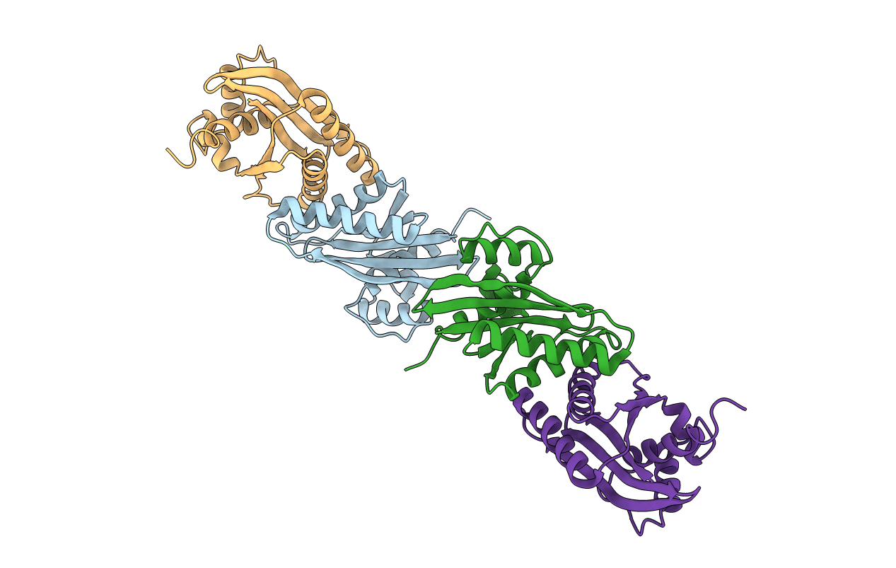
ATOMIC STRUCTURE OF THE RUVC RESOLVASE: A HOLLIDAY JUNCTION-SPECIFIC ENDONUCLEASE FROM E. COLI
Biological Source:
Source Organism:
Escherichia coli (Taxon ID: 562)
Method Details:
Experimental Method:
Resolution:
2.50 Å
R-Value Work:
0.15
R-Value Observed:
0.15
Space Group:
P 1 21 1
Macromolecular Entities
Polymer Type:polypeptide(L)
Molecule:HOLLIDAY JUNCTION RESOLVASE (RUVC)
Gene (Uniprot):ruvC
Chain IDs:A, B, C, D
Chain Length:158
Number of Molecules:4
Biological Source:Escherichia coli
Primary Citation
Atomic structure of the RuvC resolvase: a holliday junction-specific endonuclease from E. coli.
Cell(Cambridge,Mass.) 78 1063 1072 (1994)
PMID: 7923356 DOI: 10.1016/0092-8674(94)90280-1

Abstact

The crystal structure of the RuvC protein, a Holliday junction resolvase from E. coli, has been determined at 2.5 A resolution. The enzyme forms a dimer of 19 kDa subunits related by a dyad axis. Together with results from extensive mutational analyses, the refined structure reveals that the catalytic center, comprising four acidic residues, lies at the bottom of a cleft that nicely fits a DNA duplex. The structural features of the dimer, with a 30 A spacing between the two catalytic centers, provide a substantially defined image of the Holliday junction architecture. The folding topology in the vicinity of the catalytic site exhibits a striking similarity to that of RNAase H1 from E. coli.

Legend

Protein

Chemical

Disease

Primary Citation of related structures