1HJQ image
Deposition Date 2003-02-27
Release Date 2003-07-25
Last Version Date 2024-10-23
Entry Detail
PDB ID:
1HJQ
Keywords:
Title:
Structure of two fungal beta-1,4-galactanases: searching for the basis for temperature and pH optimum.
Biological Source:
Source Organism:
Host Organism:
Method Details:
Experimental Method:
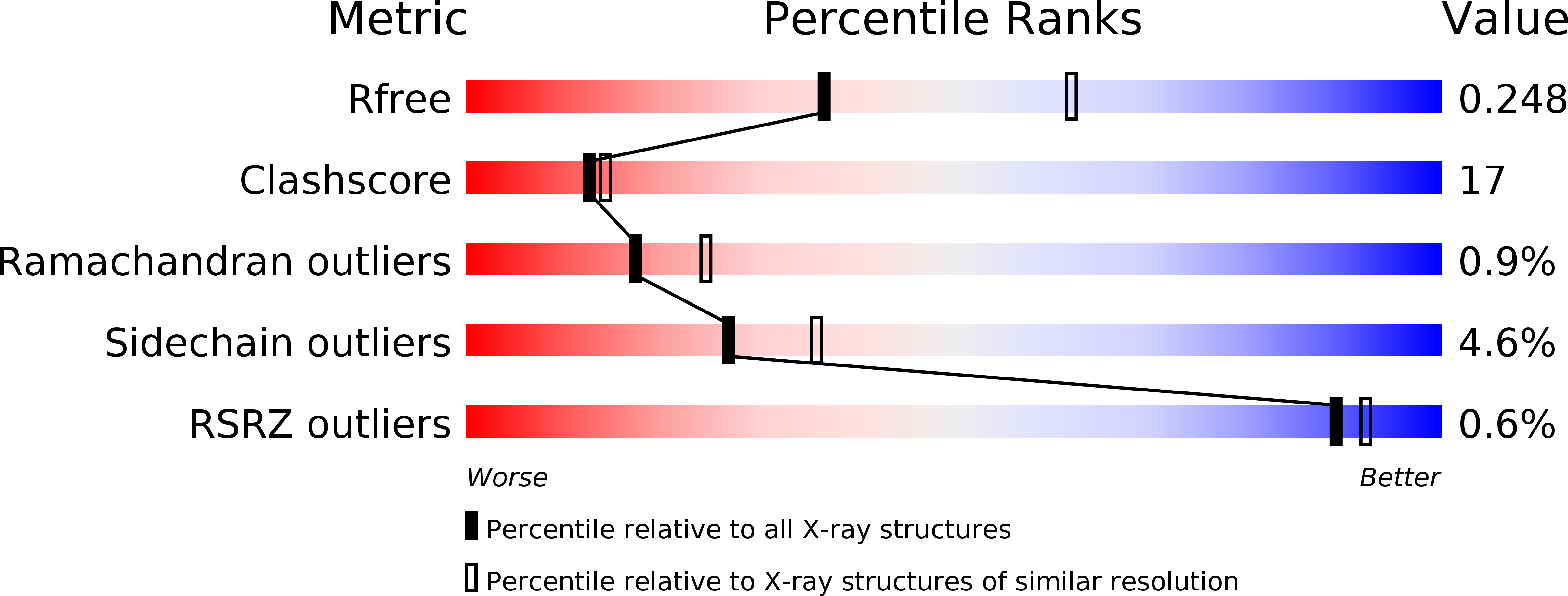
Resolution:
2.55 Å
R-Value Free:
0.24
R-Value Work:
0.19
R-Value Observed:
0.19
Space Group:
P 21 21 21
Macromolecular Entities
Polymer Type:polypeptide(L)
Molecule:BETA-1,4-GALACTANASE
Chain IDs:A
Chain Length:332
Number of Molecules:1
Biological Source:HUMICOLA INSOLENS
Modified Residue
Compound ID Chain ID Parent Comp ID Details 2D Image
ASN A ASN GLYCOSYLATION SITE
Ligand Molecules
Primary Citation
Structure of Two Fungal Beta-1,4-Galactanases: Searching for the Basis for Temperature and Ph Optimum
Protein Sci. 12 1195 ? (2003)
PMID: 12761390 DOI: 10.1110/PS.0300103

Abstact

beta-1,4-Galactanases hydrolyze the galactan side chains that are part of the complex carbohydrate structure of the pectin. They are assigned to family 53 of the glycoside hydrolases and display significant variations in their pH and temperature optimum and stability. Two fungal beta-1,4-galactanases from Myceliophthora thermophila and Humicola insolens have been cloned and heterologously expressed, and the crystal structures of the gene products were determined. The structures are compared to the previously only known family 53 structure of the galactanase from Aspergillus aculeatus (AAGAL) showing approximately 56% identity. The M. thermophila and H. insolens galactanases are thermophilic enzymes and are most active at neutral to basic pH, whereas AAGAL is mesophilic and most active at acidic pH. The structure of the M. thermophila galactanase (MTGAL) was determined from crystals obtained with HEPES and TRIS buffers to 1.88 A and 2.14 A resolution, respectively. The structure of the H. insolens galactanase (HIGAL) was determined to 2.55 A resolution. The thermostability of MTGAL and HIGAL correlates with increase in the protein rigidity and electrostatic interactions, stabilization of the alpha-helices, and a tighter packing. An inspection of the active sites in the three enzymes identifies several amino acid substitutions that could explain the variation in pH optimum. Examination of the activity as a function of pH for the D182N mutant of AAGAL and the A90S/ H91D mutant of MTGAL showed that the difference in pH optimum between AAGAL and MTGAL is at least partially associated with differences in the nature of residues at positions 182, 90, and/or 91.

Legend

Protein

Chemical

Disease

Primary Citation of related structures