1HJ9 image
Deposition Date 2001-01-10
Release Date 2002-01-04
Last Version Date 2025-04-09
Entry Detail
PDB ID:
1HJ9
Keywords:
Title:
Atomic resolution structures of trypsin provide insight into structural radiation damage
Biological Source:
Source Organism:
BOS TAURUS (Taxon ID: 9913)
Method Details:
Experimental Method:
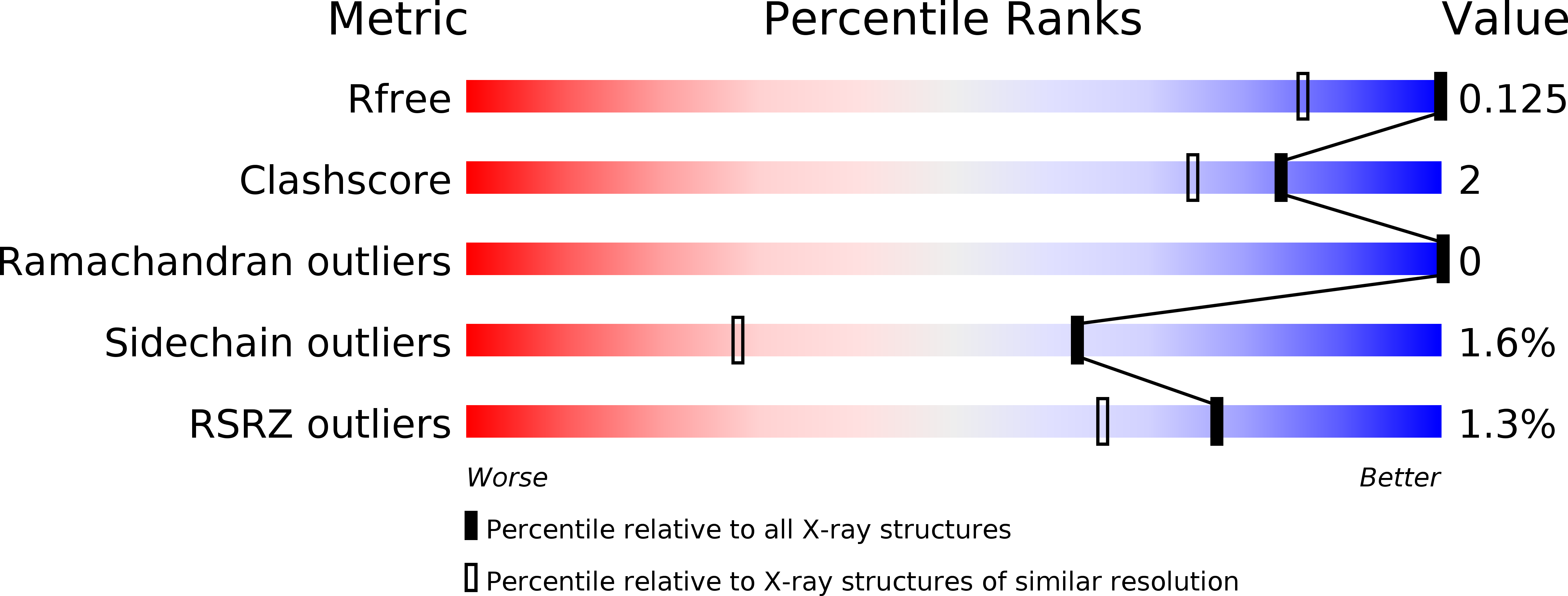
Resolution:
0.95 Å
R-Value Free:
0.13
R-Value Observed:
0.11
Space Group:
P 21 21 21
Macromolecular Entities
Polymer Type:polypeptide(L)
Molecule:BETA-TRYPSIN
Gene (Uniprot):PRSS1
Chain IDs:A
Chain Length:223
Number of Molecules:1
Biological Source:BOS TAURUS
Primary Citation
Atomic Resolution Structure of Trypsin Provide Insight Into Structural Radiation Damage
Acta Crystallogr.,Sect.D 57 488 ? (2001)
PMID: 11264577 DOI: 10.1107/S0907444901000646

Abstact

Radiation damage is an inherent problem in protein X-ray crystallography and the process has recently been shown to be highly specific, exhibiting features such as cleavage of disulfide bonds, decarboxylation of acidic residues, increase in atomic B factors and increase in unit-cell volume. Reported here are two trypsin structures at atomic resolution (1.00 and 0.95 A), the data for which were collected at a third-generation synchrotron (ESRF) at two different beamlines. Both trypsin structures exhibit broken disulfide bonds; in particular, the bond from Cys191 to Cys220 is very sensitive to synchrotron radiation. The data set collected at the most intense beamline (ID14-EH4) shows increased structural radiation damage in terms of lower occupancies for cysteine residues, more breakage in the six disulfide bonds and more alternate conformations. It appears that high intensity and not only the total X-ray dose is most harmful to protein crystals.

Legend

Protein

Chemical

Disease