Deposition Date
1992-02-12
Release Date
1993-10-31
Last Version Date
2024-10-23
Entry Detail
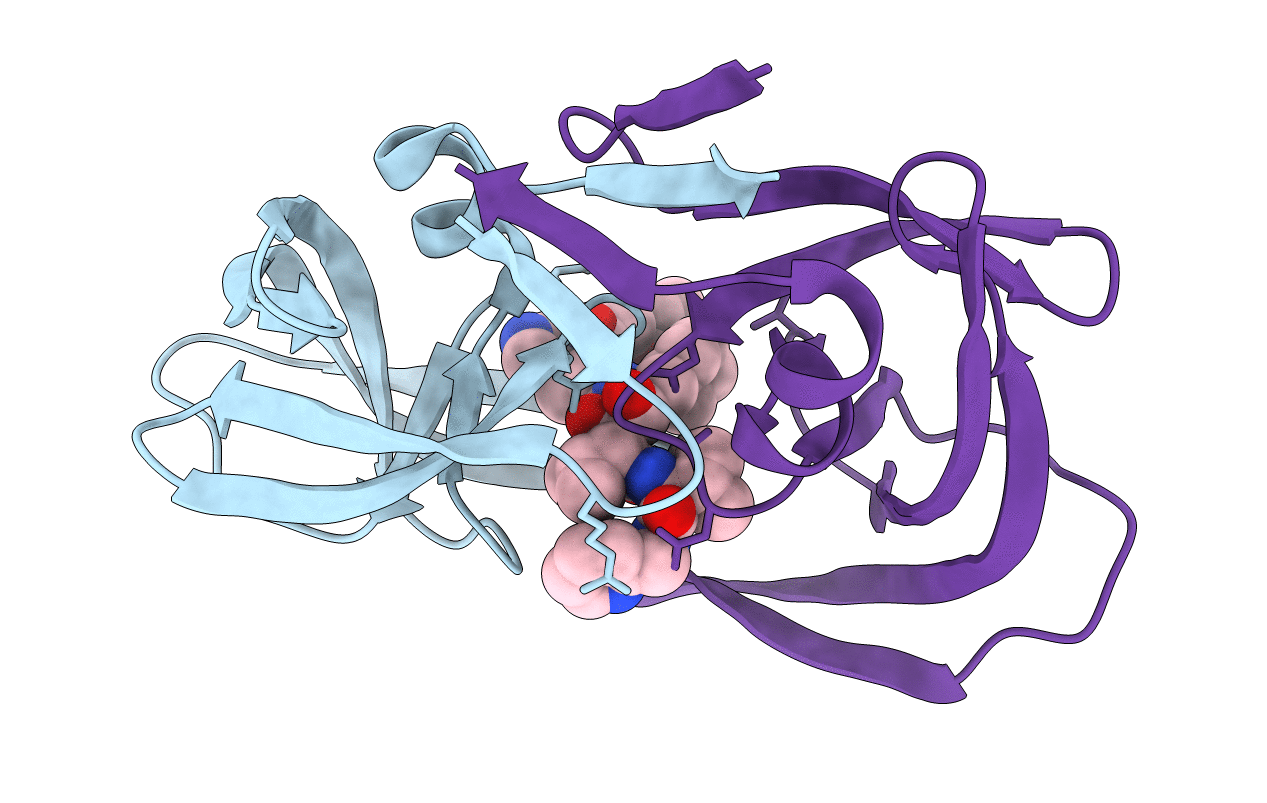
PDB ID:
1HIV
Keywords:
Title:
CRYSTAL STRUCTURE OF A COMPLEX OF HIV-1 PROTEASE WITH A DIHYDROETHYLENE-CONTAINING INHIBITOR: COMPARISONS WITH MOLECULAR MODELING
Biological Source:
Source Organism(s):
Human immunodeficiency virus 1 (Taxon ID: 11676)
Expression System(s):
Method Details:
Experimental Method:
Resolution:
2.00 Å
R-Value Observed:
0.16
Space Group:
P 21 21 21