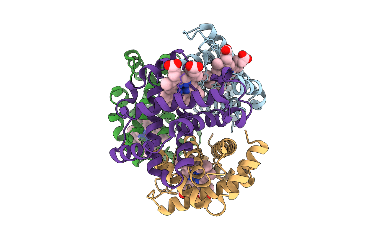
1HGA image
Entry Detail
PDB ID:
1HGA
Title:
HIGH RESOLUTION CRYSTAL STRUCTURES AND COMPARISONS OF T STATE DEOXYHAEMOGLOBIN AND TWO LIGANDED T-STATE HAEMOGLOBINS: T(ALPHA-OXY)HAEMOGLOBIN AND T(MET)HAEMOGLOBIN
Biological Source:
Source Organism:
PDB Version:
Deposition Date:
1991-10-31
Release Date:
1994-01-31
Method Details:
Experimental Method:
Resolution:
2.10 Å
R-Value Observed:
0.20
Space Group:
P 21 21 2
Macromolecular Entities
Polymer Type:polypeptide(L)
Description:HEMOGLOBIN (DEOXY) (ALPHA CHAIN)
Chain IDs:A, C
Chain Length:141
Number of Molecules:2
Biological Source:Homo sapiens
Polymer Type:polypeptide(L)
Description:HEMOGLOBIN (DEOXY) (BETA CHAIN)
Chain IDs:B, D
Chain Length:146
Number of Molecules:2
Biological Source:Homo sapiens
Ligand Molecules
Primary Citation
High resolution crystal structures and comparisons of T-state deoxyhaemoglobin and two liganded T-state haemoglobins: T(alpha-oxy)haemoglobin and T(met)haemoglobin.
J.Mol.Biol. 228 551 579 (1992)
PMID: 1453464 DOI: 10.1016/0022-2836(92)90842-8

Abstact

The origin of co-operativity in haemoglobin (Hb) resides in the reduced affinity of the T-state. T-state Hb crystals grown from polyethyleneglycol can be liganded without the molecule switching to the R high affinity state. X-ray analysis of T-state alpha-oxy Hb and T-state met Hb has identified the structural basis for reduced affinity. The nature of the chemical tension at the haem environment is different in the alpha and beta haems. There are small but definite structural changes associated with ligation in the T-state: these prove to be mostly in the same direction as the larger changes that occur in the T-->R transition.

Legend

Protein

Chemical

Disease

Primary Citation of related structures