1HCI image
Deposition Date 2001-05-04
Release Date 2001-06-27
Last Version Date 2023-12-13
Entry Detail
PDB ID:
1HCI
Title:
CRYSTAL STRUCTURE OF THE ROD DOMAIN OF ALPHA-ACTININ
Biological Source:
Source Organism:
HOMO SAPIENS (Taxon ID: 9606)
Host Organism:
Method Details:
Experimental Method:
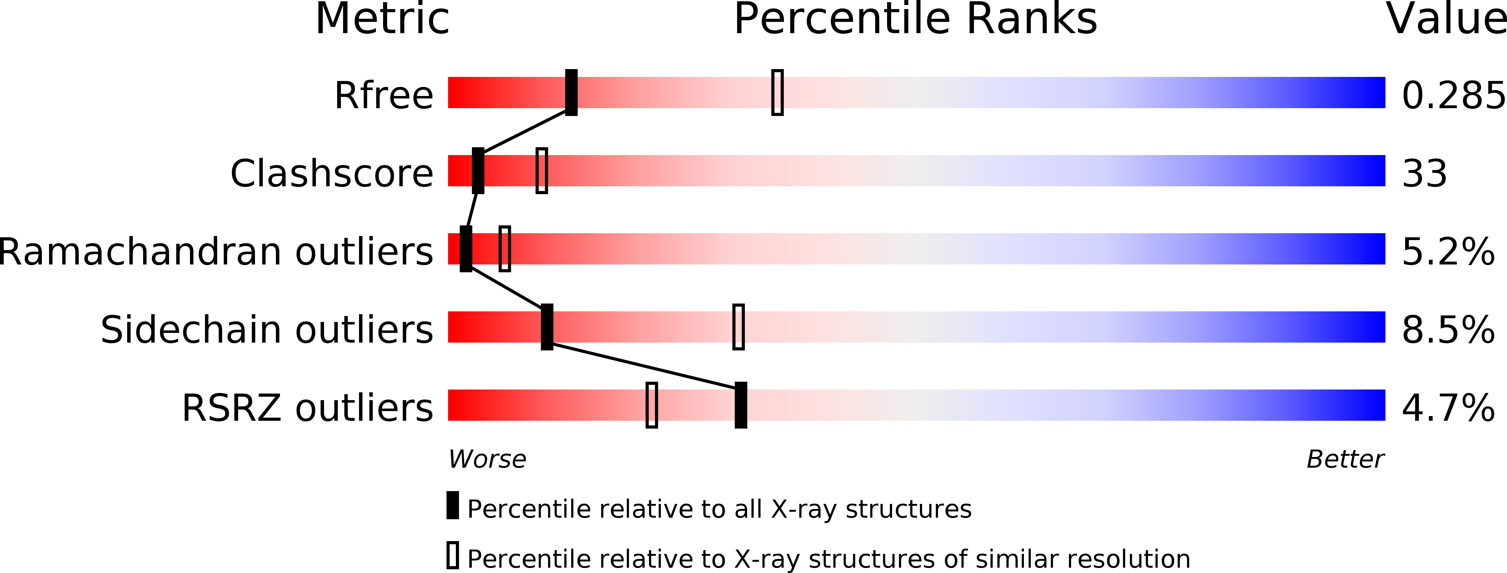
Resolution:
2.80 Å
R-Value Free:
0.29
R-Value Work:
0.27
R-Value Observed:
0.27
Space Group:
P 31
Macromolecular Entities
Polymer Type:polypeptide(L)
Molecule:ALPHA-ACTININ 2
Gene (Uniprot):ACTN2
Chain IDs:A, B
Chain Length:476
Number of Molecules:2
Biological Source:HOMO SAPIENS
Primary Citation
Crystal Structure of the Alpha-Actinin Rod Reveals an Extensive Torsional Twist
Structure 9 597 ? (2001)
PMID: 11470434 DOI: 10.1016/S0969-2126(01)00619-0

Abstact

BACKGROUND: Alpha-actinin is a ubiquitously expressed protein found in numerous actin structures. It consists of an N-terminal actin binding domain, a central rod domain, and a C-terminal domain and functions as a homodimer to cross-link actin filaments. The rod domain determines the distance between cross-linked actin filaments and also serves as an interaction site for several cytoskeletal and signaling proteins. RESULTS: We report here the crystal structure of the alpha-actinin rod. The structure is a twisted antiparallel dimer that contains a conserved acidic surface. CONCLUSIONS: The novel features revealed by the structure allow prediction of the orientation of parallel and antiparallel cross-linked actin filaments in relation to alpha-actinin. The conserved acidic surface is a possible interaction site for several cytoplasmic tails of transmembrane proteins involved in the recruitment of alpha-actinin to the plasma membrane.

Legend

Protein

Chemical

Disease

Primary Citation of related structures