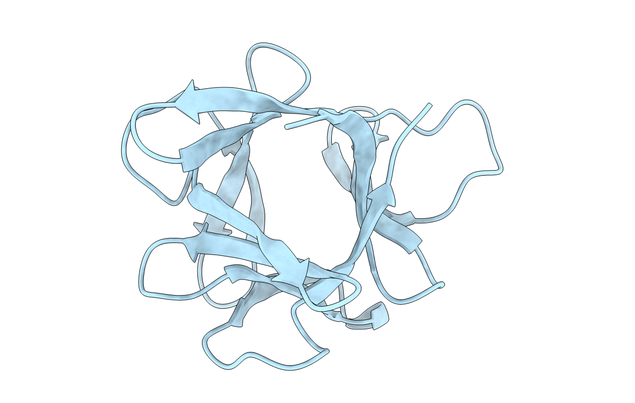
1HCD image
Entry Detail
PDB ID:
1HCD
Keywords:
Title:
STRUCTURE OF HISACTOPHILIN IS SIMILAR TO INTERLEUKIN-1 BETA AND FIBROBLAST GROWTH FACTOR
Biological Source:
Source Organism:
PDB Version:
Deposition Date:
1994-05-03
Release Date:
1994-10-15
Method Details:
Experimental Method:
Conformers Submitted:
1
Macromolecular Entities
Polymer Type:polypeptide(L)
Description:HISACTOPHILIN
Chain IDs:A
Chain Length:118
Number of Molecules:1
Biological Source:Dictyostelium discoideum
Ligand Molecules
Primary Citation
Structure of hisactophilin is similar to interleukin-1 beta and fibroblast growth factor.
Nature 359 855 858 (1992)
PMID: 1436061 DOI: 10.1038/359855a0

Abstact

The fast reaction of the actin-based cytoskeleton in motile cells after stimulation with a chemoattractant requires a signal-transduction chain that creates a very specific environment at distinct regions beneath the plasma membrane. Dictyostelium hisactophilin, a unique actin-binding protein, is a submembranous pH sensor that signals slight changes of the H+ concentration to actin by inducing actin polymerization and binding to microfilaments only at pH values below seven. It has a relative molecular mass of 13.5K and its most unusual feature is the presence of 31 histidine residues among its total of 118 amino acids. The transduction of an external signal from the plasma membrane to the cytoskeleton is poorly understood. Here we report the protein's structure in solution determined by nuclear magnetic resonance spectroscopy. The nuclear Overhauser effect intensities of the three-dimensional nuclear Overhauser spectra were used directly in the calculations. The overall folding of histactophilin is similar to that of interleukin-1 beta and fibroblast growth factor, but the primary amino-acid sequence of hisactophilin is unrelated to these two proteins.

Legend

Protein

Chemical

Disease

Primary Citation of related structures